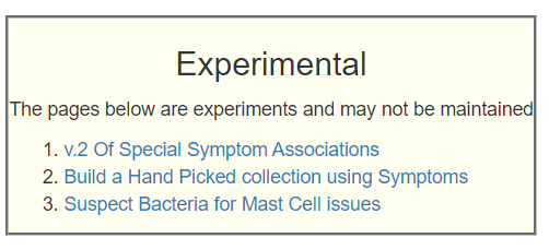
Microbiome: What goes in the mouth, comes out the other end!

I have written about this in the past:

When I first started Microbiome Prescription the main and most popular provider was uBiome (dearly departed) which explicitly offered oral microbiome analysis. I had about a dozen uploads. Eventually I stopped support because there was not going to be sufficient uploads, even if I was waited for a decade.

Stomach Acid does not nuke probiotics!

This is a common internet legend which disagrees with both studies and common sense!!!

Common Sense Anyone?

This is the key question: “If you have a bacteria in your gut, how did it get there?”

Assuming that you do not believe that bacteria has perfected teleportation, then there is just one route: Through the mouth, the stomach, etc. It is the great trek!

The trek that every bacteria (or ancestor) in your microbiome must make (with some side trips!). Image from Wikipedia.

Studies Supporting No or Little Impact

There are two German probiotic that are suspended in water, added to water and drunk. These studies indicate that they do survive! In fact, they persist!

Looking at Vet Practices, adding probiotic to food or water is well establish. Some sites actually advocates opening capsules. Custom Probiotics advocates all of their probiotics be taken in a glass of water.

https://www.customprobiotics.com/mm5/merchant.mvc?Screen=PROD&Product_Code=CP-2024

Where does this myth come from?

I strongly suspect marketing — if you compare the cost per BCFU from Custom Probiotics to your usual health food store probiotic capsules you will see the costs can be 10x higher in the capsules That is NOT the cost of putting them into capsules. In marketing, claims about the importance of capsules is differentiate product to get you to buy brand X and not Y.

People are also willing to pay for convenience. Buying flour to bake a load of bread is much cheaper than buying a ready made load of bread. At our local coop, a custom loaf of bread was selling for $11.95 — people are lazy.

Vendor Distractor?

I also suspect that stomach acid eliminating probiotic has been used as an excuse by many vendors. The likely cause of probiotics not staying around is where they were sourced: Human or Animal. “In general, their optimal growth temperature ranges between 36–38°C and 41–43°C for human and animal origin strains,” [2011] A probiotic bacteria is unlikely to thrive at a temperature 7°C (or 12°F) from it’s preferred temperature. It will likely reproduce less and be less robust (allowing other bacteria to beat it up).

This comes back to my old soapbox: Buy Only Probiotics where the STRAIN (and not just the species!) is listed – and that specific strain has been researched (Ideally for the condition that concerns you. Use this link to look up most, for example periodontal disease), and that the origin is human. I would suggest constantly emailing the manufacturers!!! It is likely the only way that the situation will improve.

Oral Microbiome Is associated with many conditions

My impression is that any condition with a neurological component (i.e. brain fog, impulse control, etc) is likely to have ORAL microbiome dysfunction.

Going Forward

There are many products available, for example, see this list. They will likely contain one or more of the following. For a bigger list, see researched probiotics here: mouth, sinus and oral.

  • Bacillus Coagulans
  • BLIS K12,
  • BLIS M18,
  • lactobacillus acidophilus,
  • lactobacillus reuteri,
  • lactobacillus paracasei,
  • lactobacillus salivarius,
  • salivarius thermophilus
  • Symbioflor-1 [2012] Enterococcus faecalis

The “salivarius” indicate where it was first identified (mouth saliva). So it is normally in the mouth.

The key is for the probiotic to stay in the mouth for sufficient time to dislodge some other residents. To me, this appears to suggest:

  • Brush and rinse after every meal
  • You could do things like break apart an Oregano Oil capsule or drop Monolaurin flakes into the mouth and hold them there for as long as you can tolerate them…
  • Take one or more lozenge afterwards… I would suggest taking one at a time, then perhaps change to a different one when dissolved. There are a few probiotics that are available as pressed pills. If the taste is not too bad, I personally use Miyarisan(jp) [clostridium butyricum] and shin biofermin (jp) [Bifidobacterium bifidum, Enterococcus faecalis, Lactobacillus acidophilus] in this manner. Some others to consider:
    • BioGaia Prodentis Mint Lozenges [Lactobacillus Reuteri]
    • Flora, Super 5 Probiotic Lozenges [Lactobacillus Acidophilus: 60% Bifidobacterium Bifidum: 15% Lactobacillus Bulgaricus: 15% Streptococcus Thermophilus: 5% Lactobacillus Salivarius: 5%]
    • NOW Supplements, OralBiotic™, [Streptococcus salivarius BLIS K12]

The ideal would be to take an oral microbiome test that reports percentile ranking of each bacteria (against other oral samples). I do not know of any one providing that. uBiome likely had that data, but they are no more.

Sudden Illness Triggered ME/CFS

For more Analysis Posts on Long COVID and ME/CFS click here.

Back Story

I’m male, 5’ 10”, currently 145 lbs, 60 years old. I first became ill suddenly in 1987, with what at the time seemed to be food poisoning or a stomach bug. The nausea, stomach upset and loss of appetite lasted months after, I became bedridden. Dozens of tests, doctors, revealed nothing.

Gradually over time, the symptoms subsided and I began to eat and gain weight again. After about a year and a half, I became functional, but never recovered to my previous state. This has been the course of life since. The symptoms would reoccur, last several months, then subside. With no definitive cause for beginning, nor treatment for ending.

The ongoing fatigue over the years was relentless. I somehow managed to complete a 30 year carrier, and took retirement at first opportunity. Doctors speculated that my work was a stress factor responsible for my condition, and retirement would solve it. It didn’t.

For the past several years, I’m mostly housebound, able to go outside and do minor tasks on occasion. Currently, my worst of symptoms are LPR/ reflux related. Not in the traditional sense, mine is a gas/ aero, that I believe is being caused by severe dysbiosis/ imbalance.

I cite this study as an example,

Accompanied with voice loss, throat and chest pain, severe at times.

A recent endoscopy showed “mild gastritis”. Doctors offer me benzodiazepines and antidepressants, stating my symptoms do not correlate with their findings.

Previous endoscopes/ colonoscopies were unremarkable. Gastric empty test normal.

I have tested negative for SIBO several times. IBS Smart test, h pylori, celiac, mast cell (MCAS), all negative.

Numerous diets, eliminations, supplements, herbs, prescribed medications have brought no help or relief. Most have made symptoms worse.

I did manage to have a biopsy taken and sent to Dr John Chia, to test for entero virus. It came back highly positive, however, I am somewhat skeptical that it could be a red herring. My vague attempts with pre/ probiotics resulted with increased gas and/ or diarrhea.

One clue, on two occasions (1995, 2014) after having colonoscopy, I mysteriously had remissions afterwards, that lasted several months. The speculation, is that the prep somehow created a reset of bacteria/ flora. I recently tried to replicate, by doing a prep cleanse. However, despite drinking a full gallon plus, I ran out of solution before being completely cleared out. I felt a brief improvement, but have suffered with horrid lower gas/ flatulence since.

Not the result I was after.

Initial Comments on Back Story

For myself, stress was the trigger of each of my ME/CFS episode so the speculation by his MDs was reasonable. The 1987 event, and the resulting cascade of the microbiome is the root concern. The microbiome evolves, just like society evolves. In 1987, most homes had a VCR and a few lucky people had digital pagers. Today, very few have VCRs (in use) and almost every one has a smart cellular phone. In 2023, arguing whether the right choice should be BetaMax or VHS has become irrelevant. Similarly, focusing on the cause in 1987 is really irrelevant. It may have been a virus, Lyme disease or a dozen other culprits – it is very unlikely to be relevant to addressing today’s microbiome.

Analysis

Looking at the distribution by frequency, we see an over-population of bacteria with low levels.

The Bacteria over 90% and Bacteria under 10% are a simple statistic to understand. If you have 188 different genus and true randomness then you would expect around 19 in each group. We has 12 over 90%, close, but a whopping 61 under 10% — that 32% of all bacteria, not 10%!!! In other words, we have a massive number of different bacteria at low levels. It is not a problem of a few bacteria being too high.

PercentileGenusSpecies
0 – 96176
10 – 192016
20 – 291416
30 – 3998
40 – 491520
50 – 591414
60 – 691620
70 – 791022
80 – 891722
90 – 1001221

Looking at Dr. Jason Hawrelak Recommendations for levels, he was at the 99.7%ile and the very few misses for being ideal.. they were border line.. (i.e. 15.1 versus 15; 0 versus 0.0001). In short, almost an ideal microbiome by that criteria.

Going Forward

Doing the usual 3 suggestions sets, we

The list of top suggestions look very close to what I was taking for my own remission (pre-microbiome analysis days).

  • rosmarinus officinalis (rosemary)
  • thyme (thymol, thyme oil)
  • peppermint (spice, oil)
  • lactobacillus paracasei (probiotics)
  • lemongrass oil
  • Human milk oligosaccharides (prebiotic, Holigos, Stachyose)
  • neem
  • syzygium aromaticum (clove)
  • Sumac(Rhus coriaria)
  • cinnamon (oil. spice)
  • Ajwain (trachyspermum ammi)
  • Curcumin
  • lactobacillus casei (probiotics)
  • thiamine hydrochloride (vitamin B1)
  • aloe vera
  • ashwagandha (withania somnifera)
  • garlic (allium sativum)
  • olea europaea (olive leaf)

Looking at the to-avoid

  • vsl#3 (probiotics)
  • walnuts
  • lactulose
  • amaranth
  • barley, oat
  • arabinoxylan oligosaccharides (prebiotic)
  • fish oil
  • saccharomyces cerevisiae (probiotics)
  • low protein diet : typically seen when B-vitamins are wanted.

The full details are below

Going over to the Food Site we see:

  • Chicken and Turkey Liver being at the top of the list
mg per 100 gramsSignificant amountNutrient
25800SignificantProtein
13.925SignificantNiacin
12.9SignificantIron, Fe
4.293SignificantRetinol
2.313SignificantRiboflavin
0.84SignificantPyridoxine (Vitamin B6
0.56SignificantFolic acid
0.02113SignificantVitamin B-12
The key nutrient components typically from Livers

Pass 2 — Looking at Prescription items

One non-prescription showed up near the top, an item that I have used with good effect: monolaurin. The top antibiotics list include:

The above have various risks, and should be review carefully. My own preferences would be minocycline first, then hyoscyamine [because IBS is a factor for this patient]. I should note that using a different algorithm without consensus (Special Reports for your MDs) reports contrary results.

Feedback on Antibiotics

Another freak incident that resulted in a five month remission. In 2011, I had one tonsil that became huge. The other remained completely normal. Doctors were suspect of cancer, and both were removed. Thankfully, it was not cancer. So they didn’t look any further to determine the cause.

But at 49 year old, this was no picnic. Rough surgery and recovery, but it followed with probably the most significant remission of all. It was an amazing turn around, all symptoms backed off, energy returned to nearly normal. After 5 months though, symptoms began to return, and within the year I was back to my previous state

I was on amoxicillin for several weeks after the surgery. When the symptoms began to return, we became suspect that it was the amoxicillin that had done something. My doctor put me back on it, but there was no improvement. Whatever had taken place, was a random-chance occurrence. Maybe the amoxicillin was responsible, by creating a shift in bacteria balance.

I found that Cecil Jadin’s protocol is what I tend to advocate. One of the key reason is that it was it was tuned from many years of clinical experience at the Pasteur Institute for Tropical Medicine for what they termed as an “Occult Rickettsia” infection. The basis of it is rotating different families of antibiotics. The mathematics are simple — first round may eliminate 90% of the issues with 10% being resistant. A different antibiotic usually require a different type of resistance, so 90% of the remaining 10% is eliminated.. leaving just 1%. Doing a third round, takes us down to 0.1%

I speculate that the few survivors from your first round of amoxicillin slowly rebuild . Because these bacteria were the survivors, those with resistance genes. The repopulated gut was largely resistant, hence no effect from this antibiotic later. Think of it as a boxing match. You landed a good punch — but instead of landing more punches, the opponent was able to recover and block the same punch later.

Later, discussion of this story with a well-meaning gastrologist, he had me do a week of xifaxan. He felt certain, based on my story, it would get me back to the previous gains. It made me horrid sick, lasting for weeks after stopping it. There was no improvement.

My preference is not to pick antibiotics by symptoms (or what worked for the prior patient), but from the bacteria results that are desired. Results are not guaranteed — rather, IMHO this approach has higher probability of being successful.

Looking at Probiotics

First, using KEGG data: as is typical for most ME/CFS people (and consistent with the ME/CFS conference reports from 1998): Escherichia coli (Symbioflor-2 or Mutaflor). The next common one is Bacillus subtilis (natto), Clostridium butyricum, Lacticaseibacillus casei, Enterococcus faecalis.

From the consensus list we see a good overlap and have in order:

bacillus subtilis natto is the source for a supplement called nattokinase, which dissolves fibrin deposits and also an anti-inflammatory [2021]. It is also available not as a probiotic, but in a Japanese Dessert Food called Natto. Natto is an acquired taste.

Natto: Available in some Japanese groceries stores

My probiotic suggestions would be the following (at sufficient dosage, see this page):

  • 2 weeks of Bacillus subtilis (perhaps 10 BCFU daily)
  • 2 weeks of lactobacillus casei (perhaps 48 BCFU daily)
  • 2 weeks of one of the E.Coli probiotics (Mutaflor: 4 capsules per day, starting at 1 and slowly increasing)

Then repeat. Note that some probiotics are strong avoid, for example: saccharomyces cerevisiae / saccharomyces boulardii.

Taking Herbs

There are a large number of herbs cited above. In keeping with my philosophy of avoiding resistance, take some of herbs for 2 weeks and then change to others herbs for the next two weeks. The question is how to take it? I know that some will claim that tinctures are more effective; IMHO, tinctures are very effective for reducing back accounts!

My personal practice is to take herbs in one of two ways:

  • Buying them in bulk, organic and making our own 000 capsules. Most store purchased herb capsules do not appear to be organic, often with additional ingredients “to make them better” – which is often marketing hype.
    • We take them immediately prior to meals so that the stomach acid produced to handle the meal, also dissolves the active ingredients from the herbs.
  • Taking them as a hot tea. Some herbs are horrible tasting… those tend to end up as capsules.

Many, but not all, herbs have documented dosages with links to studies (which can be informative for how to take). For example: Neem: 120 mg/day, Olive Leaf: 700 mg/day, Curcumin: 3 gm/day. My general rule of thumb is one 000 capsule with each meal.

Questions

Q: Curious to know, do you think there may be an advantage of using this method with probiotics, to deliver past the stomach, farther down the gut?

A: I know this common belief, but have not seen any clinical studies demonstrating it. What I have seen is probiotics delivered as a liquid in water, are documented to persist for weeks after a single dose. That is, the specific strain delivered was not detected before but was detected in subsequent weeks. This indicates that this belief is very questionable. Personally, I tend to use single documented strains of probiotics from Custom Probiotics and follow their directions. I do keep food at least a hour away from taking probiotics so stomach acid production will be quiet.

https://www.customprobiotics.com/l-casei-probiotic-powder.html

On a related issue, remember that the gut is downstream from the mouth and nasal passages. The source of bad bacteria may be there and may account for repopulation over time. One probiotic that has been shown effective for the nasal passages etc is Symbioflor-1. There are a few hard tablet probiotics out there (for example, Miyarisan — Clostridium butyricum). I have often just put them in my mouth and let them dissolve there.

NOTE: I will be doing a follow up post on The oral microbiome, coming soon!

An Exercise showing the Volatility of Bacteria Counts

This post started out seeking to confirm or debunk the claim located here.

The method was very simple because we have a continuous stream of samples from before COVID, before the COVID vaccination and after the majority of people uploading samples would have been vaccinated. If this massive change is happening then the pre-COVID bifidobacterium count (by lab) would be much higher than the post-COVID vaccination bifidobacterium counts.

My results: there was no statistical significance between the averages

  • Pre 2020-01-01: Average Count 20380 on 118 samples, Std Dev 98300
  • Post 2022-06-01: Average Count 26111 on 406 samples, Std Dev 72700

That is a 28% increase when a decrease was expected from the above talk.

I am open data, so you can pull the data and check the calculations:

Volatility of Numbers

I was also curious to see if there was any apparent month by month pattern, so I pulled the statistics for biidobacterium, shown below. It is illuminating to a statistician like me, perhaps confusing or concerning to people with poor understanding of statistics (who would expect the numbers from month to month to be similar).

ThryveBiomeSight
YearMonthAverageStd DevObsAverageStd DevObs
202073243813164624279293582614
20208254564340521768389489
20209134101932917185012256614
20201084056148144184370739020
202011185983404994926819713
2020121007817108161841271829
202116815217240520124362067532
2021210160016398030172895550945
202135795710324817144823377433
2021421979429673077002443646
20215246935174456142573360838
20216281668449139214658576251
202174702310520939226206722951
20218602838239843184277978437
20219624389292928120021963541
2021101312129924245922856538
20211111515270955799962496658
202112285828019117114982991963
2022115114287603870761514950
20222248165906932117072720252
20223104862399533202435153947
2022410207215805779161828869
20225334718249780103042371981
20226238616012653805321994235
202272679710943540107092043962
20228677071321086080851719085
20229139261762228126352133292
2022109090140494596272117189
20221110296190343972931215061
2022123186619421108872139042
20231102241821541133022161289
2023210604228835291031978172
20233659537817330135663425657
Statistics for Bifidobacterium

My conclusion is that you need to have two things to get good results:

  • All of the samples should be processed by the same lab at the same time. Different batches of reagents may cause different results.
  • You need good sample sizes, at least 100+
  • You need to be very very careful not to cherry pick data (example below)

An example from Thryve/Ombre data above, with a sample size of 30, the average was 101600. Later a sample size of 21 reported just 3186. Conclusion: going back to school caused family bifidobacterium to tank!

On sample size of 100 issue:

Improving Iron Levels

This is an update of my 2016 post: Low Iron – A Gut Bacteria Connection. One key addition is that Vitamin A supplementation may have significant positive impact.

The microbiota shifts the iron sensing of intestinal cells [2016]. “The amount of iron in the diet directly influences the composition of the microbiota. Inversely, the effects of the microbiota on iron homeostasis have been little studied….Commensal organisms (Bacteroides thetaiotaomicron VPI-5482 and Faecalibacterium prausnitzii A2-165) and a probiotic strain (Streptococcus thermophilus LMD-9) led to up to 12-fold induction of ferritin in colon.”

Probiotics to Take

Likely No or Negative Effect

Biomesight vs Thorne Tests – Differences

This is a person as in this prior set of posts, A Microbiome Trek Continues thru the land of ME/CFS.

There are two key differences that needs to be understood

  • Difference in numbers reported (of bacteria and percentiles)
  • Will the suggestions change?

Comparison of Results: Thorne to Biomesight

The samples were only a few weeks apart, so similar data. The ratio of bacteria reporting is 6x more for Thorne than Biomesight, so the expectation would be similar shifts for most of the others.

Key Difference: Bacteria Percentiles come from Thorne, the other percentiles are computed against a composite of other samples (until we get sufficient samples). The Conditions, Enzymes and Compound estimates are likely unreliable (we compare against all tests and not other samples from the same procession) and we will ignore in this analysis.

Many of the criteria are identical between tests: Outside Range for JasonH, Medivere, Metagenomics , MyBioma, Nirvana/CosmosId and XenoGene. So for people using those criteria — there is no difference between the tests.

The Bacteria over 90% and Bacteria under 10% are a simple statistic to understand. 10% should be under 10% and 10% above the 90%ile to have a balance microbiome.

With Thorne we have 3226 bacteria and true randomness then you would expect around 322 in each group. We find 239 over 90%, close, but a whopping 1577 under 10% — that 48% of all bacteria, not 10%!!! In other words, we have a massive number of different bacteria at low levels. It is not a problem of a few bacteria being too high (which is a common belief about gut dysfunction), but many only have token amounts.

For Biomesight, we have 503, and thus would expect 50 and 50. For over 90%ile, we have just 25, and for under 10%, 108 bacteria. The high %ile is just 50% of expected and 200% of expected for low with Biomesight; Thorne is just 75% of expected for high, but a massive 489% of expected for low.

CriteriaThorne SampleBiomesight
Bacteria Reported By Lab3226503
Bacteria Over 99%ile19310
Bacteria Over 95%ile21218
Bacteria Over 90%ile23925
Bacteria Under 10%ile1577108
Bacteria Under 5%ile141144
Bacteria Under 1%ile11063
Different Labs – Items Skipped
Pathogens16234
Outside Range from JasonH66
Outside Range from Medivere1616
Outside Range from Metagenomics77
Outside Range from MyBioma55
Outside Range from Nirvana/CosmosId1717
Outside Range from XenoGene3535
Outside Lab Range (+/- 1.96SD)1898
Outside Box-Plot-Whiskers68527
Outside Kaltoft-Moldrup175391
Condition Est. Over 99%ile66
Condition Est. Over 95%ile157
Condition Est. Over 90%ile2410
Enzymes Over 99%ile9310
Enzymes Over 95%ile673118
Enzymes Over 90%ile1131647
Enzymes Under 10%ile312150
Enzymes Under 5%ile26275
Enzymes Under 1%ile18312
Compounds Over 99%ile230100
Compounds Over 95%ile498463
Compounds Over 90%ile684606
Compounds Under 10%ile1350599
Compounds Under 5%ile1336580
Compounds Under 1%ile1324569

Comparison of Results: Thorne to Ombre

The Bacteria over 90% and Bacteria under 10% are a simple statistic to understand. If you have 3226 bacteria and true randomness then you would expect around 322 in each group.

  • For Ombre we would expect 59 over 90%ile and under 10%ile. close. We have 22 or 37% of expected for low %ile and 117 or 200% of expected for low percentile.

Many of the criteria are identical between tests: Outside Range for JasonH, Medivere, Metagenomics , MyBioma, Nirvana/CosmosId and XenoGene. So for people using those criteria — there is no difference between the tests.

CriteriaThorne SampleOmbre
Bacteria Reported By Lab3226588
Bacteria Over 99%ile1932
Bacteria Over 95%ile21210
Bacteria Over 90%ile23922
Bacteria Under 10%ile1577117
Bacteria Under 5%ile141167
Bacteria Under 1%ile11066
Different Labs – Items Skipped
Pathogens16234
Outside Range from JasonH77
Outside Range from Medivere1414
Outside Range from Metagenomics55
Outside Range from MyBioma88
Outside Range from Nirvana/CosmosId1818
Outside Range from XenoGene4646
Outside Lab Range (+/- 1.96SD)1895
Outside Box-Plot-Whiskers68534
Outside Kaltoft-Moldrup1753129
Condition Est. Over 99%ile60
Condition Est. Over 95%ile150
Condition Est. Over 90%ile240
Enzymes Over 99%ile930
Enzymes Over 95%ile6739
Enzymes Over 90%ile1131101
Enzymes Under 10%ile312165
Enzymes Under 5%ile26265
Enzymes Under 1%ile1832
Compounds Over 99%ile23038
Compounds Over 95%ile498236
Compounds Over 90%ile684332
Compounds Under 10%ile1350248
Compounds Under 5%ile1336159
Compounds Under 1%ile132433

My personal opinion is that Thorne is better because the more bacteria reported, the greater the statistical significance of over and under representation. On the positive side, all three samples agree on the shifts of bacteria patterns

Analysis

The distribution continues to match a common pattern with ME/CFS microbiomes, an over abundance of low percentile bacteria. This is also seen with the prior Biomesight sample. This shift is made much stronger with Thorne because more genus and species are reported. It also emphasis the shifts seen above.

PercentileGenusSpecies
0 – 9417628
10 – 198582
20 – 296085
30 – 394269
40 – 493432
50 – 5959339
60 – 692660
70 – 791628
80 – 891531
90 – 9921142
Thorne Report
PercentileGenusSpecies
0 – 92433
10 – 193237
20 – 291523
30 – 39911
40 – 49916
50 – 59814
60 – 6989
70 – 791017
80 – 89715
90 – 99610
Biomesight Report

Treatment Dilemma

The usual algorithm is to increase bacteria with low percentiles and decrease those with high percentiles. When you have a huge numbers of low percentile then the question arises: Do you really want to increase these, or do you want to eliminate them entirely to get them off the radar? It is a valid question, but to do that, we have to make increase/eliminate suggestions on 417 genus. That is not practical (given the sparseness of data and limited knowledge of so many genus). My working hypothesis is that keeping to the usual algorithm is the best way to go. Let the bacteria make the determination of winners and losers.

Going Forward

I am going to build two consensus reports. One for Thorne and one for the latest Biomesight, then use the “Uber Consensus” report on the Multiple Samples tab. The purpose is to see whether there are really significant differences in suggestions between the two sample reports.

We are going to do 4 basic suggestions for each:

The results had 581 suggestions. I did a Pivot tables of Take Counts against Avoid Counts to visualize the similarities between each set of suggestions going into the uber suggestions. I read the pivot table below as indicating that the suggestions were equivalent with 73 items being to Take with no Avoid, and a further 72 items with just 1 avoid. We have lots of choices in agreement

CountsTakes
Avoid012345678
045162311122
1323711672
233111019529
3206911547
41518942911
512101441
632251
737
813

What are some of the top suggestions?

As with the prior reports, Escherichia coli probiotics is at the top of the KEGG computed probiotics, typical for ME/CFS. From the consensus, we have:

The absence of most Lactobacillus is not surprising because they are hostile to Escherichia Coli. My pivot conference report from 1998 had this bacteria being low in ME/CFS patients. With Thorne, we can get actual numbers (16s numbers for Escherichia Coli are questionable). This person Thorne Results is at the 27%ile for Escherichia and 29%ile for Escherichia Coli, which is consistent with that conference report and the KEGG computation for probiotics.

Out of interest, I looked for the %ile on the Thorne results of the consensus suggested probiotics:

Having the actual percentiles for the strains used in probiotics allows us to tune the suggestions. In this case, we should skip any probiotics with bacillus subtilis or clostridium butyricum. There is no point in taking them. On the other side, you have confirmation that the suggested probiotics are likely to have an impact. I give the Thorne results a big 👍 because you can actually determine the probiotics that you likely not benefit from. The 16s results only report a few probiotic species (with questionable accuracy).

The Extras from the Thorne Results

This person did not see the next data on Thorne’s Web Site — but it was in the data CSV file to upload. The virus count with a few having percentiles. The ones without percentiles are rare ones without data.

Virus

And Fungi too!

For both of these sets of data, values over 90% should be researched. Fungi are of special concern because they can both be treated often and may also indicate a mold issue around the person.

User Feedback

Thanks Ken! .. and yikes . Mold is my nemesis. I’ve been trying to figure if I’ve had mold / moldy house for years. I did an ermi for the entire house and got a 2 which is really low. Recently I did an air test all around the house and it was pretty low minus a car which I knew was an issue and I’ve been trying to avoid. I’m trying to decipher if it’s a past issue and I can’t detox or current 🧐🧐

Attached is my air test.

Question: yellow highlights on Thorne. Those were 90%+ , there were some non highlighted 90% + results. Should I only research the ones you highlighted?

I should have a new mold urine test result coming too which I’ll send your way once I get it!

Answers: As a general and very rough rule, count the number of items reported (Virus: 35, Fungi:50). Take this number and multiple by a percentile – say 99%ile, and round up. For Virus and Fungi it is 1. This is the number of false positives that would be expected with 99% or higher.

You appear to have an issue. Your mold test fortunately identifies the genus and where it is located. The HVAC and washing machine hints that it is brought into the house, likely on clothes. The attic appears free of the ones that you are high in. I noticed that Malassezia is not reported in the mold test.

Bottom Line

IMHO, getting a Thorne sample is a definite should do at least once. Why? some of your issues may be fungi or virus related. The difference for suggestions of using Thorne, BiomeSight or Ombre is slight. The differences are reasonable given the sparseness of the data that we have for suggestions.

Postscript – and Reminder

I am not a licensed medical professional and there are strict laws where I live about “appearing to practice medicine”.  I am safe when it is “academic models” and I keep to the language of science, especially statistics. I am not safe when the explanations have possible overtones of advising a patient instead of presenting data to be evaluated by a medical professional before implementing.

I cannot tell people what they should take or not take. I can inform people items that have better odds of improving their microbiome as a results on numeric calculations. I am a trained experienced statistician with appropriate degrees and professional memberships. All suggestions should be reviewed by your medical professional before starting.

The answers above describe my logic and thinking and is not intended to give advice to this person or any one. Always review with your knowledgeable medical professional.

The Dice of Health – A game of craps

My uber focus for the last few years has been on the microbiome. The reasons are simple: relatively rich amount of data to work from, detail tests can be done without a Physician’s Order, and treatment can often be done without a prescription.

In no way am I saying that the microbiome is the complete picture. It is simply the easiest to doddle in.

The analogy of a dice is good to get the entire picture. Actually two dice … because often you feel like crap as a result of a roll of the die in the craps game of life.

Some Sides of The Die

The following are the sides that come quickly into mind, they are likely more

  1. SNP/DNA issues. Many conditions have associations with specific DNA mutations.
  2. Infections (Past or Present)
  3. Environment
  4. Minerals
  5. Vitamins
  6. Organic Acid and Other Metabolites
  7. Microbiome
  8. Epigenetics

Chances are that a condition will develop when two (or more) die are rolled with bad values

Worked Example

I am using Chronic Fatigue Syndrome (CFS) / Myalgic Encephalomyelitis (ME) because I am most familar with the existing literature. The same can be done for many other conditions – for example Autism.

SNP/DNA for ME/CFS

A few examples of findings

Infections (Current or Past)

Side Note: Many cancers are associated with specific virial infections.

Environment

Minerals

This can be a function of environment, diet, water quality.

Vitamins

Organic Acid and Other Metabolites

Within this, stomach acid and blood pH is included.

Microbiome

A quick copy and paste. For many other conditions, see this page.

📓 Potential role of microbiome in Chronic Fatigue Syndrome/Myalgic Encephalomyelits (CFS/ME).
Scientific reports (Sci Rep ) Vol: 11 Issue 1 Pages: 7043
Pub: 2021 Mar 29 Epub: 2021 Mar 29 Authors Lupo GFD , Rocchetti G , Lucini L , Lorusso L , Manara E , Bertelli M , Puglisi E , Capelli E ,
Summary Html Article Publication
📓 Gut Microbiota Interventions With <i>Clostridium butyricum</i> and Norfloxacin Modulate Immune Response in Experimental Autoimmune Encephalomyelitis Mice.
Frontiers in immunology (Front Immunol ) Vol: 10 Issue Pages: 1662
Pub: 2019 Epub: 2019 Jul 23 Authors Chen H , Ma X , Liu Y , Ma L , Chen Z , Lin X , Si L , Ma X , Chen X ,
Summary Html Article Publication
📓 Correction to: Open-label pilot for treatment targeting gut dysbiosis in myalgic encephalomyelitis/chronic fatigue syndrome: neuropsychological symptoms and sex comparisons.
Journal of translational medicine (J Transl Med ) Vol: 16 Issue 1 Pages: 39
Pub: 2018 Feb 23 Epub: 2018 Feb 23 Authors Wallis A , Ball M , Butt H , Lewis DP , McKechnie S , Paull P , Jaa-Kwee A , Bruck D ,
Summary Html Article Publication
📓 Potential role of dengue virus, chikungunya virus and Zika virus in neurological diseases.
Memorias do Instituto Oswaldo Cruz (Mem Inst Oswaldo Cruz ) Vol: 113 Issue 11 Pages: e170538
Pub: 2018 Oct 29 Epub: 2018 Oct 29 Authors Vieira MADCES , Costa CHN , Linhares ADC , Borba AS , Henriques DF , Silva EVPD , Tavares FN , Batista FMA , Guimarães HCL , Martins LC , Monteiro TAF , Cruz ACR , Azevedo RDSDS , Vasconcelos PFDC ,
Summary Html Article Publication
📓 Human Gut-Derived Commensal Bacteria Suppress CNS Inflammatory and Demyelinating Disease.
Cell reports (Cell Rep ) Vol: 20 Issue 6 Pages: 1269-1277
Pub: 2017 Aug 8 Epub: Authors Mangalam A , Shahi SK , Luckey D , Karau M , Marietta E , Luo N , Choung RS , Ju J , Sompallae R , Gibson-Corley K , Patel R , Rodriguez M , David C , Taneja V , Murray J ,
Summary Html Article Publication
📓 Fecal metagenomic profiles in subgroups of patients with myalgic encephalomyelitis/chronic fatigue syndrome.
Microbiome (Microbiome ) Vol: 5 Issue 1 Pages: 44
Pub: 2017 Apr 26 Epub: 2017 Apr 26 Authors Nagy-Szakal D , Williams BL , Mishra N , Che X , Lee B , Bateman L , Klimas NG , Komaroff AL , Levine S , Montoya JG , Peterson DL , Ramanan D , Jain K , Eddy ML , Hornig M , Lipkin WI ,
Summary Html Article Publication
📓 A Pair of Identical Twins Discordant for Myalgic Encephalomyelitis/Chronic Fatigue Syndrome Differ in Physiological Parameters and Gut Microbiome Composition.
The American journal of case reports (Am J Case Rep ) Vol: 17 Issue Pages: 720-729
Pub: 2016 Oct 10 Epub: 2016 Oct 10 Authors Giloteaux L , Hanson MR , Keller BA ,
Summary Html Article
📓 Support for the Microgenderome: Associations in a Human Clinical Population.
Scientific reports (Sci Rep ) Vol: 6 Issue Pages: 19171
Pub: 2016 Jan 13 Epub: 2016 Jan 13 Authors Wallis A , Butt H , Ball M , Lewis DP , Bruck D ,
Summary Html Article Publication
📓 Chronic fatigue syndrome patients have alterations in their oral microbiome composition and function.
PloS one (PLoS One ) Vol: 13 Issue 9 Pages: e0203503
Pub: 2018 Epub: 2018 Sep 11 Authors Wang T , Yu L , Xu C , Pan K , Mo M , Duan M , Zhang Y , Xiong H ,
Summary Publication Publication
📓 Gut-associated lymphoid tissue, gut microbes and susceptibility to experimental autoimmune encephalomyelitis.
Beneficial microbes (Benef Microbes ) Vol: 7 Issue 3 Pages: 363-73
Pub: 2016 Jun Epub: 2016 Feb 3 Authors Stanisavljevic S , Lukic J , Momcilovic M , Miljkovic M , Jevtic B , Kojic M , Golic N , Mostarica Stojkovic M , Miljkovic D ,
Summary Publication Publication
📓 Increased d-lactic Acid intestinal bacteria in patients with chronic fatigue syndrome.
In vivo (Athens, Greece) (In Vivo ) Vol: 23 Issue 4 Pages: 621-8
Pub: 2009 Jul-Aug Epub: Authors Sheedy JR , Wettenhall RE , Scanlon D , Gooley PR , Lewis DP , McGregor N , Stapleton DI , Butt HL , DE Meirleir KL ,
Summary

Epigenetics

This is where an event, like stress, causes the behavior of DNA to change. Your DNA is the same, just a “switch” is turned on or off.

Going Forward with Treatment

My attitude is evidence based action with testable models. If you walk into a physician’s office, it is unlikely that they will be aware with the many sides of the dice. Usually, they want simple “follow the recipe book” cases where what to do is clear.

For myself, I had the luxury of unbelievable, unlimited, medical coverage for a few years. I found some of the DNA issues, and to quote a physician “You are extremely lucky with that mutation, it is very treatable” — I became a piracetam addict when needed. Most people do not have that luxury.

Looking at 8 items above, I ask the same question:

  • Is it objective measurable?
    • Can you get the test (willing MD, cost)
  • Is it treatable?
    • Do we have actual clinical studies showing treatment is effective?
      • Is the treatment just symptom relief or remission?
    • What are the risk of side-effects?

If getting information from a test is not clearly actionable, then it does not help with treatment and not worth the expense. Testing for testing sake is a luxury for the rich.

My Criteria in evaluating new proposed models. “

Many people will advocate that just one of these 8 sides of the die needs to be done for a cure. IMHO, if the model does not address most of these factors, it is likely to work for only a few.

For me, the Microbiome model appears the best to use.

  • Microbiome tests are cheap and do not require a MD to be involved — Objective
  • We have hundreds of studies showing substances alters the microbiome
  • Risk of side-effects with non-prescription items is low

And it is connected to the other factors above well.

  • Many of the organic acid and metabolites are produced by the microbiome. Thus correcting the microbiome is likely to resolve this I compute many of these using Kyoto Encyclopedia of Genes and Genomes data.
  • Vitamins and Mineral absorption is deeply influences by the microbiome too!

If you have DNA information, for example on your methylation, this impacts your microbiome and the reverse. Being tested for DNA SNPs that does not have effective treatment is a waste of money. The individual’s microbiome is greatly influenced by their DNA. They co-exist and co-operate. In some cases, the microbiome bacteria can produce anticoagulants and fibrinolytics which can counter some coagulation issues.

WARNING ON PEOPLE PROPOSING MODELS

Over the last 30 years, I have constantly seen people proposing this model or that model. Usually the model is focusing on a single aspect of one the die sides above. For ME/CFS, it was the search for an occult virus that was the root cause of this condition. This often comes out of a need to reduce to the simple in whatever specialty that the researcher or physician is trained in. The wages of over-specialization in modern medicine. Be wary of any model that does not offer a concrete explanation for all of the laboratory results in the literature. Often models will cherry-pick studies and ignore the majority of other studies, or do vague hand waving.

The cause is almost never just one of the above factors, but typically many.

Some Feedback from using Microbiome Prescription

Recently I have been getting several emails from people with status updates. I thought that I should share a few of them. I have not gotten any negative feedback (Are all of my readers Canadians who are too polite to complain?)

Also good news. I’ve managed to correct much of my microbiome. I’ve reversed the NIH gnavus ans prausnitzii signature that is very common in ME. My prausnitzii is now 21%! It was something like 0.2 two years ago.

And my lactobacillus and bifido are in the very bottom range of healthy for the first time in two years. Also three lactobacillus strains from vivomixx appeared on my 16s for the first time. Proving that even artificial probiotics can populate the gut albeit temporarily. 

I intend to continue and get another test in three months. While my physical symptoms have improved a lot my light sensitivity and brainfog are still not great to be honest and I have no idea why. Leaky gut can I think be ruled out becuass gnavus is so low and I barely react to eggs anymore (I’ve had issues with eggs since I was a kid). But I have some work to do still.

I really love the website and get a ton of use out of it.  I hope you stick with it and keep updating it and making it better.

Mold is what got me initially too.  Retrospectively.  When we moved to our new house is when my decline actually started.  And then when we redid our master bath, there was a bunch of mold, and I got real sick real fast.

In essence, I had all the triggers.  Every single one of these:

  1. Viral or bacterial exposure (listed in order of severity) – COVID and RSV
  2. Trauma – to intestines
  3. Food poisoning – Bacterial and fungal
  4. Prolonged Stress – Luxonis.com startup, I’m the founder
  5. Environmental Toxins – Mold in our MASTER BEDROOM

Oh forgot to mention I took lactobacillus Rhamnosus based my my research before I noticed your big red note to not take it and other lactobacillus because they block the impact of heparin.  I think that’s what really got me!

Haven’t pooped for 3 days since that mistake!  Before that pooped every day for 14. 

As an update, I’m nearly 5 weeks in and am beginning to feel better.  My energy levels are perhaps the best they’ve been in the last 5 years.  I’ve still got a very long way to go but the results thus far are promising!

I’m taking 5-7 foods/ supplements, 2-3X a day.  And every 2 weeks I’m rotating all of it to prevent antibiotic resistance.  In another month I plan to retest myself and make the necessary adjustments to my protocol.  

The other reason I’m writing is that a friend with similar fatigue issues and a histamine intolerance has just gotten tested and is joining my journey to recovery. 

Long COVID – an update

First, there is a body of literature indicating that the microbiome plays an important part in many conditions. The last one in this list of citations agrees with my hypothesis for the last 7 years: “Multi-kingdom gut microbiota analyses define CONDITION-X severity and in some case the syndrome.

Citation
📓 Reversion of Gut Microbiota during the Recovery Phase in Patients with Asymptomatic or Mild COVID-19: Longitudinal Study.
Microorganisms (Microorganisms ) Vol: 9 Issue 6 Pages:
Pub: 2021 Jun 7 Epub: 2021 Jun 7 Authors Kim HN , Joo EJ , Lee CW , Ahn KS , Kim HL , Park DI , Park SK ,
Summary Html Article Publication
📓 The gut microbiome of COVID-19 recovered patients returns to uninfected status in a minority-dominated United States cohort.
Gut microbes (Gut Microbes ) Vol: 13 Issue 1 Pages: 1-15
Pub: 2021 Jan-Dec Epub: Authors Newsome RC , Gauthier J , Hernandez MC , Abraham GE , Robinson TO , Williams HB , Sloan M , Owings A , Laird H , Christian T , Pride Y , Wilson KJ , Hasan M , Parker A , Senitko M , Glover SC , Gharaibeh RZ , Jobin C ,
Summary Html Article Publication
📓 Gut Microbiota May Not Be Fully Restored in Recovered COVID-19 Patients After 3-Month Recovery.
Frontiers in nutrition (Front Nutr ) Vol: 8 Issue Pages: 638825
Pub: 2021 Epub: 2021 May 13 Authors Tian Y , Sun KY , Meng TQ , Ye Z , Guo SM , Li ZM , Xiong CL , Yin Y , Li HG , Zhou LQ ,
Summary Html Article Publication
📓 Gut Microbiota Interplay With COVID-19 Reveals Links to Host Lipid Metabolism Among Middle Eastern Populations.
Frontiers in microbiology (Front Microbiol ) Vol: 12 Issue Pages: 761067
Pub: 2021 Epub: 2021 Nov 5 Authors Al Bataineh MT , Henschel A , Mousa M , Daou M , Waasia F , Kannout H , Khalili M , Kayasseh MA , Alkhajeh A , Uddin M , Alkaabi N , Tay GK , Feng SF , Yousef AF , Alsafar HS ,
Summary Publication
📓 Gut microbiota dynamics in a prospective cohort of patients with post-acute COVID-19 syndrome.
Gut (Gut ) Vol: Issue Pages:
Pub: 2022 Jan 26 Epub: 2022 Jan 26 Authors Liu Q , Mak JWY , Su Q , Yeoh YK , Lui GC , Ng SSS , Zhang F , Li AYL , Lu W , Hui DS , Chan PK , Chan FKL , Ng SC ,
Summary Publication
📓 Alterations in microbiota of patients with COVID-19: potential mechanisms and therapeutic interventions.
Signal transduction and targeted therapy (Signal Transduct Target Ther ) Vol: 7 Issue 1 Pages: 143
Pub: 2022 Apr 29 Epub: 2022 Apr 29 Authors Wang B , Zhang L , Wang Y , Dai T , Qin Z , Zhou F , Zhang L ,
Summary Publication
📓 Alterations in Gut Microbiota of Patients With COVID-19 During Time of Hospitalization.
Gastroenterology (Gastroenterology ) Vol: 159 Issue 3 Pages: 944-955.e8
Pub: 2020 Sep Epub: 2020 May 20 Authors Zuo T , Zhang F , Lui GCY , Yeoh YK , Li AYL , Zhan H , Wan Y , Chung ACK , Cheung CP , Chen N , Lai CKC , Chen Z , Tso EYK , Fung KSC , Chan V , Ling L , Joynt G , Hui DSC , Chan FKL , Chan PKS , Ng SC ,
Summary Publication
📓 Alterations in Fecal Fungal Microbiome of Patients With COVID-19 During Time of Hospitalization until Discharge.
Gastroenterology (Gastroenterology ) Vol: 159 Issue 4 Pages: 1302-1310.e5
Pub: 2020 Oct Epub: 2020 Jun 26 Authors Zuo T , Zhan H , Zhang F , Liu Q , Tso EYK , Lui GCY , Chen N , Li A , Lu W , Chan FKL , Chan PKS , Ng SC ,
Summary Publication
📓 Challenges in the Management of SARS-CoV2 Infection: The Role of Oral Bacteriotherapy as Complementary Therapeutic Strategy to Avoid the Progression of COVID-19.
Frontiers in medicine (Front Med (Lausanne) ) Vol: 7 Issue Pages: 389
Pub: 2020 Epub: 2020 Jul 7 Authors d`Ettorre G , Ceccarelli G , Marazzato M , Campagna G , Pinacchio C , Alessandri F , Ruberto F , Rossi G , Celani L , Scagnolari C , Mastropietro C , Trinchieri V , Recchia GE , Mauro V , Antonelli G , Pugliese F , Mastroianni CM ,
Summary Publication
📓 It Ain`t Over `Til It`s Over: SARS CoV-2 and Post-infectious Gastrointestinal Dysmotility.
Digestive diseases and sciences (Dig Dis Sci ) Vol: Issue Pages:
Pub: 2022 Mar 30 Epub: 2022 Mar 30 Authors Coles MJ , Masood M , Crowley MM , Hudgi A , Okereke C , Klein J ,
Summary Publication
📓 Integrated analysis of gut microbiome and host immune responses in COVID-19.
Frontiers of medicine (Front Med ) Vol: 16 Issue 2 Pages: 263-275
Pub: 2022 Apr Epub: 2022 Mar 8 Authors Xu X , Zhang W , Guo M , Xiao C , Fu Z , Yu S , Jiang L , Wang S , Ling Y , Liu F , Tan Y , Chen S ,
Summary Publication
📓 Respiratory dysfunction three months after severe COVID-19 is associated with gut microbiota alterations.
Journal of internal medicine (J Intern Med ) Vol: 291 Issue 6 Pages: 801-812
Pub: 2022 Jun Epub: 2022 Mar 17 Authors Vestad B , Ueland T , Lerum TV , Dahl TB , Holm K , Barratt-Due A , Kåsine T , Dyrhol-Riise AM , Stiksrud B , Tonby K , Hoel H , Olsen IC , Henriksen KN , Tveita A , Manotheepan R , Haugli M , Eiken R , Berg Å , Halvorsen B , Lekva T , Ranheim T , Michelsen AE , Kildal AB , Johannessen A , Thoresen L , Skudal H , Kittang BR , Olsen RB , Ystrøm CM , Skei NV , Hannula R , Aballi S , Kvåle R , Skjønsberg OH , Aukrust P , Hov JR , Trøseid M , NOR-Solidarity study group. ,
Summary Publication
📓 Dissecting the role of the human microbiome in COVID-19 via metagenome-assembled genomes.
Nature communications (Nat Commun ) Vol: 13 Issue 1 Pages: 5235
Pub: 2022 Sep 6 Epub: 2022 Sep 6 Authors Ke S , Weiss ST , Liu YY ,
Summary Publication
📓 Multi-kingdom gut microbiota analyses define COVID-19 severity and post-acute COVID-19 syndrome.
Nature communications (Nat Commun ) Vol: 13 Issue 1 Pages: 6806
Pub: 2022 Nov 10 Epub: 2022 Nov 10 Authors Liu Q , Su Q , Zhang F , Tun HM , Mak JWY , Lui GC , Ng SSS , Ching JYL , Li A , Lu W , Liu C , Cheung CP , Hui DSC , Chan PKS , Chan FKL , Ng SC ,
Summary Publication

Looking at what is statistically significant in samples uploaded to the citizen science site, we see that only BiomeSight has sufficient data. It identifies 36 taxa/bacteria at present. With more samples coming in, this may increase. Before looking at the bacteria, I like to look at the enzymes — why? different bacteria produce the same enzymes so it is an elegant way of clustering bacteria by what they produce.

KEGG Enzyme data

In this case, I am blown away on the number of statistically significant shifts! If you want more information about these enzymes, see BRENDA or/and Kyoto Encyclopedia of Genes and Genomes.

There are 197 with P < 0.001. Give there is 8000+ enzymes, that suggests around 8 may be false positives.

Enzyme NameECKEYWith
Long COVID
Without
Long
COVID
T-ScoreDFProbability
(S)-3-hydroxy-3-methylglutaryl-CoA acetoacetate-lyase (acetyl-CoA-forming)4.1.3.41303575207613.57666P < 0.001
4-amino-5-aminomethyl-2-methylpyrimidine aminohydrolase3.5.99.21516936912613.48666P < 0.001
dihydrourocanate:acceptor oxidoreductase1.3.99.331351335592113.44666P < 0.001
nucleoside-triphosphate diphosphohydrolase3.6.1.91455286352313.38666P < 0.001
poly(deoxyribonucleotide)-3′-hydroxyl:5′-phospho-poly(deoxyribonucleotide) ligase (ATP or NAD+)6.5.1.61294755177913.34666P < 0.001
poly(deoxyribonucleotide)-3′-hydroxyl:5′-phospho-poly(deoxyribonucleotide) ligase (ATP, ADP or GTP)6.5.1.71294755177913.34666P < 0.001
acetyl-CoA:kanamycin-B N6′-acetyltransferase2.3.1.821286735166213.32666P < 0.001
acetyl-CoA:2-deoxystreptamine-antibiotic N3-acetyltransferase2.3.1.811302005310413.28666P < 0.001
(1->4)-alpha-D-galacturonan reducing-end-disaccharide-lyase4.2.2.91280385098913.28666P < 0.001
alpha-maltose-6′-phosphate 6-phosphoglucohydrolase3.2.1.1221304965395113.28666P < 0.001
D-serine ammonia-lyase (pyruvate-forming)4.3.1.181285765198713.23666P < 0.001
ATP phosphohydrolase (ABC-type, iron(III) enterobactin-importing)7.2.2.171303735418013.15666P < 0.001
protein-Npi-phospho-L-histidine:D-mannose Npi-phosphotransferase2.7.1.1911407016280713.14666P < 0.001
protein-Npi-phospho-L-histidine:D-mannitol Npi-phosphotransferase2.7.1.1971292845372313.06666P < 0.001
ADP-alpha-D-glucose:alpha-D-glucose-1-phosphate 4-alpha-D-glucosyltransferase (configuration-retaining)2.4.1.3421346545802813.04665P < 0.001
aryl-ester hydrolase3.1.1.21511717237713.02666P < 0.001
ATP phosphohydrolase (ABC-type, Fe3+-transporting)7.2.2.71421526611112.86666P < 0.001
palmitoyl-CoA hydrolase3.1.2.21494997172812.83666P < 0.001
ATP:[protein]-L-tyrosine O-phosphotransferase (non-specific)2.7.10.21319255736012.75666P < 0.001
D-psicose 3-epimerase5.1.3.301434166828412.74666P < 0.001
D-tagatose 3-epimerase5.1.3.311434166828412.74666P < 0.001
penicillin amidohydrolase3.5.1.111391386616512.62666P < 0.001
D-aspartate:[beta-GlcNAc-(1->4)-Mur2Ac(oyl-L-Ala-gamma-D-Glu-L-Lys-D-Ala-D-Ala)]n ligase (ADP-forming)6.3.1.121453567026112.57666P < 0.001
2′-(5-triphosphoribosyl)-3′-dephospho-CoA:apo-[citrate (pro-3S)-lyase] 2′-(5-phosphoribosyl)-3′-dephospho-CoA-transferase2.7.7.611491967469712.46666P < 0.001
ATP:3′-dephospho-CoA 5-triphospho-alpha-D-ribosyltransferase2.4.2.521499577578112.39666P < 0.001
2,4,6/3,5-pentahydroxycyclohexanone 2-isomerase5.3.99.111462547353812.37666P < 0.001
acetyl-CoA:citrate CoA-transferase2.8.3.101503677631812.36666P < 0.001
L-aspartate:tRNAAsx ligase (AMP-forming)6.1.1.231329396172012.22666P < 0.001
4-phospho-D-erythronate:NAD+ 3-oxidoreductase1.1.1.4091340736291412.19666P < 0.001
4-phospho-D-threonate:NAD+ 3-oxidoreductase1.1.1.4081340736291412.19666P < 0.001
poly(deoxyribonucleotide)-3′-hydroxyl:5′-phospho-poly(deoxyribonucleotide) ligase (ATP)6.5.1.11438836812812.15666P < 0.001
ATP:D-erythronate 4-phosphotransferase2.7.1.2201336316269612.13666P < 0.001
ATP:D-threonate 4-phosphotransferase2.7.1.2191336316269612.13666P < 0.001
ATP:D-glycero-alpha-D-manno-heptose 7-phosphate 1-phosphotransferase2.7.1.1681561577849111.99666P < 0.001
2-lysophosphatidylcholine acylhydrolase3.1.1.51608748681411.71666P < 0.001
UDP-alpha-D-glucose:1,2-diacyl-sn-glycerol 3-alpha-D-glucosyltransferase2.4.1.3371599608724811.57666P < 0.001
N-succinyl-LL-2,6-diaminoheptanedioate amidohydrolase3.5.1.181514828284911.49666P < 0.001
acetate:holo-[citrate-(pro-3S)-lyase] ligase (AMP-forming)6.2.1.221632459049311.44666P < 0.001
D-tagatose 1,6-bisphosphate D-glyceraldehyde-3-phosphate-lyase (glycerone-phosphate-forming)4.1.2.401408147287911.44666P < 0.001
L-iditol:NAD+ 2-oxidoreductase1.1.1.1418152410988611.34666P < 0.001
alkylated-DNA glycohydrolase (releasing methyladenine and methylguanine)3.2.2.211721698902811.33666P < 0.001
oligosaccharide 6-alpha-glucohydrolase3.2.1.101616289371211.12666P < 0.001
(3S)-citryl-CoA oxaloacetate-lyase (acetyl-CoA-forming)4.1.3.3418537510500910.73666P < 0.001
S-adenosyl-L-methionine:tRNA (adenine22-N1)-methyltransferase2.1.1.2171579389281710.69666P < 0.001
S-adenosyl-L-methionine:16S rRNA (cytidine1409-2′-O)-methyltransferase2.1.1.22718368911402710.56666P < 0.001
S-adenosyl-L-methionine:23S rRNA (cytidine1920-2′-O)-methyltransferase2.1.1.22618368911402710.56666P < 0.001
sn-glycerol 3-phosphate:quinone oxidoreductase1.1.5.317706411048610.54666P < 0.001
L-glutamate:tRNAGlx ligase (AMP-forming)6.1.1.241654489740210.38666P < 0.001
D-glycero-D-manno-heptose 7-phosphate aldose-ketose-isomerase5.3.1.2821347513524810.25666P < 0.001
beta-D-glucose 1,6-phosphomutase5.4.2.62533831699338.98666P < 0.001
type II site-specific deoxyribonuclease3.1.21.42458641652668.77666P < 0.001
uroporphyrinogen-III carboxy-lyase (coproporphyrinogen-III-forming)4.1.1.372665621844178.59666P < 0.001
ATP phosphohydrolase (ABC-type, Ni2+-importing)7.2.2.11141054863678.56666P < 0.001
acetyl-CoA:oxalate CoA-transferase2.8.3.19133160801498.41666P < 0.001
5-phospho-alpha-D-ribose 1,2-cyclic phosphate 1,2-diphosphophosphohydrolase3.1.4.57134813821838.33666P < 0.001
Carboxypeptidase Taq3.4.17.19136153840748.31666P < 0.001
(2E)-2-enoyl-CoA:NADP+ 4-oxidoreductase1.3.1.34134146819358.31666P < 0.001
2′-deoxyribonucleoside 5′-monophosphate phosphohydrolase3.1.3.89136444842518.19666P < 0.001
cellobiose 2-epimerase5.1.3.112249331476858.17666P < 0.001
type III site-specific deoxyribonuclease3.1.21.52001551416578.10666P < 0.001
D-galactonate hydro-lyase (2-dehydro-3-deoxy-D-galactonate-forming)4.2.1.6138933881818.02666P < 0.001
protein-Npi-phospho-L-histidine:maltose Npi-phosphotransferase2.7.1.208138609883257.98666P < 0.001
L-aspartate:NAD(P)+ oxidoreductase (deaminating)1.4.1.212451501694717.90666P < 0.001
protein-Npi-phospho-L-histidine:sucrose Npi-phosphotransferase2.7.1.211144741960847.85666P < 0.001
protein-Npi-phospho-L-histidine:N-acetyl-D-glucosamine Npi-phosphotransferase2.7.1.1931520881039707.64666P < 0.001
N-acetyl-alpha-D-glucosaminyl-diphospho-ditrans,octacis-undecaprenol 4-epimerase5.1.3.262594341823877.64666P < 0.001
UDP-sugar sugarphosphohydrolase3.6.1.451563031089447.63666P < 0.001
rubredoxin:superoxide oxidoreductase1.15.1.21475721016997.40666P < 0.001
UDP-2-acetamido-2,6-dideoxy-L-talose:NADP+ oxidoreductase1.1.1.3672447901762967.33666P < 0.001
(4-O-methyl)-D-glucuronate—lignin ester hydrolase3.1.1.1172657631906857.31666P < 0.001
alkylated-DNA glycohydrolase (releasing methyladenine and methylguanine)3.2.2.203069512314077.30666P < 0.001
S-adenosyl-L-methionine:16S rRNA (cytosine967-C5)-methyltransferase2.1.1.1761923481439787.28666P < 0.001
ATP:molybdopterin-synthase adenylyltransferase2.7.7.802979932187887.25666P < 0.001
UDP-N-acetyl-alpha-D-glucosamine hydro-lyase (inverting; UDP-2-acetamido-2,6-dideoxy-beta-L-arabino-hex-4-ulose-forming)4.2.1.1152877172119847.24666P < 0.001
UDP-2-acetamido-2,6-dideoxy-beta-L-talose 2-epimerase5.1.3.282458571783087.23666P < 0.001
ATP phosphohydrolase (ABC-type, molybdate-importing)7.3.2.51578151119027.18666P < 0.001
sucrose:(1->4)-alpha-D-glucan 4-alpha-D-glucosyltransferase2.4.1.41480951022847.16666P < 0.001
UDP-N-acetyl-2-amino-2-deoxy-alpha-D-glucuronate:NAD+ 3-oxidoreductase1.1.1.3351722091248417.08666P < 0.001
an acyl-[acyl-carrier protein]:phosphate acyltransferase2.3.1.2741854451390907.05666P < 0.001
FMNH2:NAD(P)+ oxidoreductase1.5.1.391564781071797.02666P < 0.001
cellobiose:phosphate alpha-D-glucosyltransferase2.4.1.201657951177856.98666P < 0.001
4-beta-D-xylan xylohydrolase3.2.1.373084122336676.87666P < 0.001
crossover junction endodeoxyribonuclease3.1.21.103411162607196.82666P < 0.001
nucleoside-2′,3′-cyclic-phosphate 3′-nucleotidohydrolase3.1.4.163214642428306.82666P < 0.001
UDP-glucuronate 4-epimerase5.1.3.62806502075016.80666P < 0.001
S-adenosyl-L-methionine:precorrin-2 C20-methyltransferase2.1.1.1302932712275486.79666P < 0.001
S-adenosyl-L-methionine:cobalt-factor-II C20-methyltransferase2.1.1.1512931242274086.78666P < 0.001
dipeptidase E3.4.13.212042901495036.73666P < 0.001
GTP:alpha-D-mannose-1-phosphate guanylyltransferase2.7.7.133314622529986.69666P < 0.001
ATP:1,2-diacyl-sn-glycerol 3-phosphotransferase2.7.1.1073332912596976.61666P < 0.001
L-selenocysteine selenide-lyase (L-alanine-forming)4.4.1.163371722606226.56666P < 0.001
1,4-beta-D-mannooligosaccharide:phosphate alpha-D-mannosyltransferase2.4.1.3193004272273526.54666P < 0.001
4-O-beta-D-mannopyranosyl-N-acetyl-D-glucosamine:phosphate alpha-D-mannosyltransferase2.4.1.3203004272273526.54666P < 0.001
sortase B3.4.22.711613581193636.52666P < 0.001
cobalt-precorrin 5A acylhydrolase3.7.1.121607311189006.48666P < 0.001
6-carboxy-5,6,7,8-tetrahydropterin ammonia-lyase4.3.99.33290102533286.47666P < 0.001
precorrin-8 11,12-methylmutase5.4.99.601613591198066.46666P < 0.001
precorrin-8X 11,12-methylmutase5.4.99.611613591198066.46666P < 0.001
UDP-alpha-D-glucose:NAD+ 6-oxidoreductase1.1.1.223418772679686.45666P < 0.001
4-O-beta-D-mannopyranosyl-D-glucopyranose:phosphate alpha-D-mannosyltransferase2.4.1.2812945632215466.44666P < 0.001
O-acetyl-ADP-D-ribose carboxylesterase3.1.1.1062327551783806.39666P < 0.001
2′-deoxyribonucleoside-5′-diphosphate:thioredoxin-disulfide 2′-oxidoreductase1.17.4.13550182812246.39666P < 0.001
3′-ribonucleotide phosphohydrolase3.1.3.63032022295686.38666P < 0.001
acetyl-CoA:alkane-alpha,omega-diamine N-acetyltransferase2.3.1.573418062694666.32666P < 0.001
5,10-methylenetetrahydrofolate:glycine hydroxymethyltransferase2.1.2.13566062832416.30666P < 0.001
carbonic acid hydro-lyase (carbon-dioxide-forming)4.2.1.13212952489576.29666P < 0.001
L-isoleucine:tRNAIle ligase (AMP-forming)6.1.1.53640942922096.24666P < 0.001
DNA-6-O-methylguanine/DNA-4-O-methylthymine:[protein]-L-cysteine S-methyltransferase2.1.1.633656152939756.16666P < 0.001
NADH:hydroperoxide oxidoreductase1.11.1.263232752520456.11666P < 0.001
L-glutamate:tRNAGlu ligase (AMP-forming)6.1.1.173214962620666.08666P < 0.001
bleomycin hydrolase3.4.22.403144612431756.08666P < 0.001
L-cysteine-S-conjugate thiol-lyase (deaminating; 2-aminoprop-2-enoate-forming)4.4.1.133639632932376.08666P < 0.001
UDP-alpha-D-glucose:alpha-D-galactose-1-phosphate uridylyltransferase2.7.7.121700721296506.05666P < 0.001
peptidoglycan amidohydrolase3.5.1.283632332939645.99666P < 0.001
adenine-DNA deoxyribohydrolase (adenine-releasing)3.2.2.313551242853005.95666P < 0.001
D-phosphoglycerate 2,3-phosphomutase (2,3-diphosphoglycerate-dependent)5.4.2.113195822502085.95666P < 0.001
tRNA N6-(3-methylbut-2-en-1-yl)-adenine37:sulfur-(sulfur carrier),S-adenosyl-L-methionine C2-(methylsulfanyl)transferase2.8.4.33701153012245.93666P < 0.001
10-formyltetrahydrofolate:5′-phosphoribosylglycinamide N-formyltransferase2.1.2.23633742953585.89666P < 0.001
peptidase Do3.4.21.1072766602246375.87666P < 0.001
poly(deoxyribonucleotide)-3′-hydroxyl:5′-phospho-poly(deoxyribonucleotide) ligase (NAD+)6.5.1.23724633044125.85666P < 0.001
methionyl aminopeptidase3.4.11.183724623044265.85666P < 0.001
prenyl-diphosphate:adenine37 in tRNA prenyltransferase2.5.1.753718633039245.84666P < 0.001
ATP:pyridoxal 5′-phosphotransferase2.7.1.353290322617865.80666P < 0.001
L-rhamnose aldose-ketose-isomerase5.3.1.143073932414015.80666P < 0.001
peptide-methionine:thioredoxin-disulfide S-oxidoreductase [methionine (R)-S-oxide-forming]1.8.4.122577982039355.76666P < 0.001
deoxyribonuclease IV3.1.21.23536172874045.73666P < 0.001
alpha-L-rhamnoside rhamnohydrolase3.2.1.403180532527445.68666P < 0.001
ATP phosphohydrolase (ABC-type, Zn2+-importing)7.2.2.203312542645205.66666P < 0.001
L-rhamnulose-1-phosphate (S)-lactaldehyde-lyase (glycerone-phosphate-forming)4.1.2.193076322435905.63666P < 0.001
6-deoxy-6-sulfo-D-fructose:D-glyceraldehyde-3-phosphate glyceronetransferase2.2.1.14173335.6041P < 0.001
ATP:L-rhamnulose 1-phosphotransferase2.7.1.53192572556415.50666P < 0.001
ATP:glycerol 3-phosphotransferase2.7.1.302714602239705.50666P < 0.001
(R)-S-lactoylglutathione methylglyoxal-lyase (isomerizing; glutathione-forming)4.4.1.53413792800795.36666P < 0.001
exodeoxyribonuclease V3.1.11.52247101814065.36666P < 0.001
NAD(H) phosphohydrolase3.6.1.223358982753295.26666P < 0.001
L-alanyl-D-glutamate epimerase5.1.1.202119841680655.24666P < 0.001
CTP:2-C-methyl-D-erythritol 4-phosphate cytidylyltransferase2.7.7.603422402896195.12666P < 0.001
beta-D-fructofuranoside fructohydrolase3.2.1.262048151691064.91666P < 0.001
beta-lactam hydrolase3.5.2.63288922767294.76666P < 0.001
GTP 7,8-8,9-dihydrolase (diphosphate-forming)3.5.4.253631913101734.54666P < 0.001
D-ribulose 5-phosphate formate-lyase (L-3,4-dihydroxybutan-2-one 4-phosphate-forming)4.1.99.123631903101734.54666P < 0.001
5-amino-6-(D-ribitylamino)uracil butanedionetransferase2.5.1.783630213100464.54666P < 0.001
5-amino-6-(5-phospho-D-ribitylamino)uracil:NADP+ 1′-oxidoreductase1.1.1.1933560703027304.53666P < 0.001
2,5-diamino-6-hydroxy-4-(5-phospho-D-ribosylamino)pyrimidine 2-aminohydrolase3.5.4.263560703027304.53666P < 0.001
D-mannose-6-phosphate aldose-ketose-isomerase5.3.1.83499592980384.51666P < 0.001
5′-ribonucleotide phosphohydrolase3.1.3.53634173128184.40666P < 0.001
peptidoglycan-N-acetylglucosamine amidohydrolase3.5.1.1043340652891364.35666P < 0.001
fragilysin3.4.24.74772621374.30487P < 0.001
UTP:alpha-D-glucose-1-phosphate uridylyltransferase2.7.7.91800421468294.27666P < 0.001
adenosylcobinamide-GDP:alpha-ribazole ribazoletransferase2.7.8.263361012899804.27666P < 0.001
Fe(II):oxygen oxidoreductase ([FeO(OH)]core-producing)1.16.3.23438862956424.23666P < 0.001
D-stereospecific aminopeptidase3.4.11.191467321146084.23666P < 0.001
D-glycerate:NAD+ oxidoreductase1.1.1.293047902583954.21666P < 0.001
ATP:dTMP phosphotransferase2.7.4.91827271498044.20666P < 0.001
ATP:UDP-N-acetyl-alpha-D-glucosamine 3′-phosphotransferase2.7.1.176730431214.15664P < 0.001
L-histidinol:NAD+ oxidoreductase1.1.1.233607763121294.13666P < 0.001
queuosine34 in tRNA:acceptor oxidoreductase1.17.99.63658033187094.13666P < 0.001
L-lysine:tRNALys ligase (AMP-forming)6.1.1.63728373258564.10666P < 0.001
(R)-lactate hydro-lyase (adding N-acetyl-D-glucosamine 6-phosphate; N-acetylmuramate 6-phosphate-forming)4.2.1.1263154092680454.06666P < 0.001
CoA-[4′-phosphopantetheine]:apo-[acyl-carrier protein] 4′-pantetheinephosphotransferase2.7.8.71704651397214.06666P < 0.001
D-altronate hydro-lyase (2-dehydro-3-deoxy-D-gluconate-forming)4.2.1.73075682628544.05666P < 0.001
(6R)-6beta-hydroxy-1,4,5,6-tetrahydronicotinamide-adenine dinucleotide 6-epimerase5.1.99.63737213278863.98666P < 0.001
UTP:L-glutamine amido-ligase (ADP-forming)6.3.4.23696333245263.96666P < 0.001
beta-D-4-deoxy-Delta4-GlcAp-(1->3)-beta-D-GalNAc6S hydrolase3.2.1.1803002452583913.94666P < 0.001
D-erythro-1-(imidazol-4-yl)glycerol-3-phosphate hydro-lyase [3-(imidazol-4-yl)-2-oxopropyl-phosphate-forming]4.2.1.193617923162333.94666P < 0.001
1-(5-phospho-beta-D-ribosyl)-ATP:diphosphate phospho-alpha-D-ribosyl-transferase2.4.2.173618223162713.94666P < 0.001
2-phospho-4-(cytidine 5′-diphospho)-2-C-methyl-D-erythritol CMP-lyase (cyclizing; 2-C-methyl-D-erythritol 2,4-cyclodiphosphate-forming)4.6.1.123751993299063.93666P < 0.001
1-(5-phospho-beta-D-ribosyl)-5-[(5-phospho-beta-D-ribosylamino)methylideneamino]imidazole-4-carboxamide aldose-ketose-isomerase5.3.1.163617273162613.93666P < 0.001
[poly-N-acetyl-D-glucosaminyl-(1->4)-(N-acetyl-D-muramoylpentapeptide)]-diphosphoundecaprenol:[N-acetyl-D-glucosaminyl-(1->4)-N-acetyl-D-muramoylpentapeptide]-diphosphoundecaprenol disaccharidetransferase2.4.1.1293673313223813.93666P < 0.001
5-[(5-phospho-1-deoxy-D-ribulos-1-ylamino)methylideneamino]-1-(5-phospho-beta-D-ribosyl)imidazole-4-carboxamide D-erythro-1-(imidazol-4-yl)glycerol 3-phosphate-lyase (L-glutamine-hydrolysing; 5-amino-1-(5-phospho-beta-D-ribosyl)imidazole-4-carboxamide-forming)4.3.2.103631903179253.91666P < 0.001
dTDP-alpha-D-glucose 4,6-hydro-lyase (dTDP-4-dehydro-6-deoxy-alpha-D-glucose-forming)4.2.1.463633853183893.90666P < 0.001
ATP:(d)CMP phosphotransferase2.7.4.253701173258013.88666P < 0.001
L-histidinol-phosphate:2-oxoglutarate aminotransferase2.6.1.93721173279343.85666P < 0.001
5,10-methylenetetrahydrofolate,FADH2:dUMP C-methyltransferase2.1.1.1481436851151123.81666P < 0.001
orotidine-5′-phosphate carboxy-lyase (UMP-forming)4.1.1.233768413330513.81666P < 0.001
N2-formyl-N1-(5-phospho-D-ribosyl)glycinamide:L-glutamine amido-ligase (ADP-forming)6.3.5.33766903329923.80666P < 0.001
alpha-D-galactoside galactohydrolase3.2.1.223518693092623.80666P < 0.001
oligonucleotidase3.1.13.33610853184013.79666P < 0.001
ATP:pseudouridine 5′-phosphotransferase2.7.1.831431921148313.78666P < 0.001
citrate(isocitrate) hydro-lyase (cis-aconitate-forming)4.2.1.33703443270043.78666P < 0.001
adenosine-3′(2′),5′-bisphosphate 3′(2′)-phosphohydrolase3.1.3.73634183213453.74666P < 0.001
adenylyl-molybdopterin:molybdate molybdate transferase (AMP-forming)2.10.1.11558621283093.71666P < 0.001
sn-glycerol-3-phosphate:NAD(P)+ 2-oxidoreductase1.1.1.943704383286043.70666P < 0.001
isocitrate:NADP+ oxidoreductase (decarboxylating)1.1.1.423612303193823.70666P < 0.001
short-chain acyl-CoA:electron-transfer flavoprotein 2,3-oxidoreductase1.3.8.11409721132313.70666P < 0.001
acetyl-CoA:oxaloacetate C-acetyltransferase [thioester-hydrolysing, (pro-S)-carboxymethyl-forming]2.3.3.13625153211953.65666P < 0.001
UDP-2,4-diacetamido-2,4,6-trideoxy-beta-L-altropyranose hydrolase3.6.1.57560120563.64644P < 0.001
D-mannonate hydro-lyase (2-dehydro-3-deoxy-D-gluconate-forming)4.2.1.83294392881353.63666P < 0.001
ribonuclease M53.1.26.81468131199613.60666P < 0.001
(S)-4-hydroxymandelate:oxygen 1-oxidoreductase1.1.3.461483081221833.58666P < 0.001
S-adenosyl-L-methionine:tRNA (carboxymethyluridine34-5-O)-methyltransferase2.1.1.22986343.5087P < 0.001
5,10-methylenetetrahydrofolate:tRNA (uracil54-C5)-methyltransferase2.1.1.741460471200063.48666P < 0.001
aldehyde:ferredoxin oxidoreductase1.2.7.51400991144743.47666P < 0.001
cobalt-precorrin-6B:NAD+ oxidoreductase1.3.1.1061501011251203.41666P < 0.001
precorrin-6B:NADP+ oxidoreductase1.3.1.541501011251203.41666P < 0.001
(3S)-3-hydroxyacyl-CoA hydro-lyase4.2.1.171462591213433.40666P < 0.001
coproporphyrinogen-III:S-adenosyl-L-methionine oxidoreductase (decarboxylating)1.3.98.31835671567503.31666P < 0.001

Looking at the weaker statistical significance (where we expect 80 to be false positive) we have just 87 items. In other words, the ones below should likely be excluded from any analysis

Enzyme NameECKEYWith Long COVIDWithout
Long
COVID
T-ScoreDFProbability
protein-Npi-phospho-L-histidine:D-glucose Npi-phosphotransferase2.7.1.1991503951260463.30666P < 0.01
ATP:4-methyl-5-(2-hydroxyethyl)thiazole 2-phosphotransferase2.7.1.501893601640623.30666P < 0.01
D-glucose-6-phosphate:NAD+ oxidoreductase1.1.1.361368883.26359P < 0.01
ATP:alpha-D-Man-(1->2)-alpha-D-Man-(1->2)-[alpha-D-Man-(1->3)-alpha-D-Man-(1->3)-alpha-D-Man-(1->2)-alpha-D-Man-(1->2)]n-alpha-D-Man-(1->3)-alpha-D-Man-(1->3)-alpha-D-Man-(1->3)-alpha-D-GlcNAc-diphospho-ditrans,octacis-undecaprenol 3-phosphotransferase2.7.1.181386913.26346P < 0.01
S-adenosyl-L-methionine:3-O-phospho-alpha-D-Man-(1->2)-alpha-D-Man-(1->2)-[alpha-D-Man-(1->3)-alpha-D-Man-(1->3)-alpha-D-Man-(1->2)-alpha-D-Man-(1->2)]n-alpha-D-Man-(1->3)-alpha-D-Man-(1->3)-alpha-D-Man-(1->3)-alpha-D-GlcNAc-diphospho-ditrans,octacis-undecaprenol 3-phospho-methyltransferase2.1.1.294386913.26346P < 0.01
S-adenosyl-L-methionine:23S rRNA (guanine745-N1)-methyltransferase2.1.1.1871477221232113.25666P < 0.01
kanosamine-6-phosphate phosphohydrolase3.1.3.92364883.25363P < 0.01
kanosamine 6-phosphate:2-oxoglutarate aminotransferase2.6.1.104368923.24365P < 0.01
ATP:L-glutamine N5-phosphotransferase2.7.3.13241333.2354P < 0.01
GDP-beta-L-fucose:beta-D-Gal-(1->3)-alpha-D-GalNAc-(1->3)-alpha-D-GalNAc-diphospho-ditrans,octacis-undecaprenol alpha-1,2-fucosyltransferase2.4.1.308390893.22326P < 0.01
UDP-alpha-D-galactose:N-acetyl-alpha-D-galactosaminyl-R beta-1,3-galactosyltransferase (configuration-inverting)2.4.1.122390893.22326P < 0.01
UDP-alpha-D-glucose:N-acetyl-alpha-D-glucosaminyl-diphospho-ditrans,octacis-undecaprenol beta-1,3-glucosyltransferase2.4.1.305390893.22326P < 0.01
UDP-N-acetyl-alpha-D-galactosamine:N-acetyl-alpha-D-galactosaminyl-diphospho-ditrans,octacis-undecaprenol alpha-1,3-N-acetyl-D-galactosyltransferase2.4.1.306390893.22326P < 0.01
fluoroacetyl-CoA hydrolase3.1.2.292428212110223.20666P < 0.01
GDP-alpha-D-mannose:alpha-D-Man-(1->2)-alpha-D-Man-(1->2)-[alpha-D-Man-(1->3)-alpha-D-Man-(1->3)-alpha-D-Man-(1->2)-alpha-D-Man-(1->2)]n-alpha-D-Man-(1->3)-alpha-D-Man-(1->3)-alpha-D-Man-(1->3)-alpha-D-GlcNAc-diphospho-ditrans,octacis-undecaprenol 2,3-alpha-mannosyltransferase (configuration-retaining)2.4.1.371387973.20347P < 0.01
GDP-alpha-D-mannose:alpha-D-mannosyl-(1->3)-N-acetyl-alpha-D-glucosaminyl-diphospho-ditrans,octacis-undecaprenol 3-alpha-mannosyltransferase (configuration-retaining)2.4.1.349387973.20347P < 0.01
carbamoyl-phosphate:L-ornithine carbamoyltransferase2.1.3.31936661690293.19666P < 0.01
ATP:L-homoserine O-phosphotransferase2.7.1.391694241444043.19666P < 0.01
GDP-alpha-D-mannose:N-acetyl-alpha-D-glucosaminyl-diphospho-ditrans,octacis-undecaprenol 3-alpha-mannosyltransferase (configuration-retaining)2.4.1.348386983.18346P < 0.01
16S rRNA-uridine516 uracil mutase5.4.99.191912281672103.17666P < 0.01
2,3-dihydroxybenzoate:oxygen 3,4-oxidoreductase (ring-opening)1.13.11.1456243.1772P < 0.01
ATP:molybdopterin adenylyltransferase2.7.7.751509831278383.16666P < 0.01
GDP-beta-L-colitose:beta-D-galactopyranosyl-(1->3)-N-acetyl-D-glucosamine L-colitosyltransferase (configuration-inverting)2.4.1.341391963.16326P < 0.01
pyrimidine-nucleoside:phosphate (2′-deoxy)-alpha-D-ribosyltransferase2.4.2.21609661377553.13666P < 0.01
ATP phosphohydrolase (ABC-type, D-galactose-importing)7.5.2.111501901266613.13666P < 0.01
protein-Npi-phospho-L-histidine:D-fructose Npi-phosphotransferase2.7.1.2021501701271513.12666P < 0.01
diphosphate phosphohydrolase3.6.1.12049021807743.10666P < 0.01
2-oxo-acid carboxy-lyase (aldehyde-forming)4.1.1.1356953.09358P < 0.01
NAD+:diphthamide-[translation elongation factor 2] N-(ADP-D-ribosyl)transferase2.4.2.363911033.08326P < 0.01
GDP-4-dehydro-alpha-D-rhamnose 3-hydro-lyase4.2.1.1683911033.08326P < 0.01
acetyl-CoA:L-glutamate N-acetyltransferase2.3.1.11982821745523.07666P < 0.01
(8S)-3′,8-cyclo-7,8-dihydroguanosine 5′-triphosphate lyase (cyclic pyranopterin phosphate-forming)4.6.1.171648161420923.06666P < 0.01
GTP 3′,8-cyclase [(8S)-3′,8-cyclo-7,8-dihydroguanosine 5′-triphosphate-forming]4.1.99.222072831801493.05666P < 0.01
ADP-L-glycero-D-manno-heptose 6-epimerase5.1.3.2033154226353.03666P < 0.01
malonyl-CoA:malonyl-CoA malonyltransferase (decarboxylating, phloroglucinol-forming)2.3.1.2535693.0347P < 0.01
N2-acetyl-L-ornithine:L-glutamate N-acetyltransferase2.3.1.351959171724693.03666P < 0.01
dTDP-3-amino-3,4,6-trideoxy-alpha-D-glucose:2-oxoglutarate aminotransferase2.6.1.10648253.0161P < 0.01
L-threonine ammonia-lyase (2-oxobutanoate-forming)4.3.1.192873082598803.01666P < 0.01
acetyl-CoA:dTDP-4-amino-4,6-dideoxy-alpha-D-glucose N-acetyltransferase2.3.1.2093911093.01326P < 0.01
acetyl-CoA:GDP-4-amino-4,6-dideoxy-alpha-D-mannose N-acetyltransferase2.3.1.2273911093.01326P < 0.01
acetyl-CoA:polysialic-acid O-acetyltransferase2.3.1.1363911093.01326P < 0.01
UDP-alpha-D-galactose:N-acetyl-alpha-D-glucosaminyl-diphospho-ditrans,octacis-undecaprenol 3-beta-galactosyltransferase (configuration-inverting)2.4.1.3033911093.01326P < 0.01
phosphoenolpyruvate:protein-L-histidine Npi-phosphotransferase2.7.3.91983271751542.99666P < 0.01
beta-D-galactopyranosyl-(1->3)-N-acetyl-D-hexosamine:phosphate galactosyltransferase2.4.1.2111572151348372.98666P < 0.01
acyl phosphoate:sn-glycerol 3-phosphate acyltransferase2.3.1.2751881801658852.97666P < 0.01
(S)-3-hydroxybutanoyl-CoA:NADP+ oxidoreductase1.1.1.1571524421306082.95666P < 0.01
prephenate:NAD+ oxidoreductase (decarboxylating)1.3.1.122092571865582.95666P < 0.01
GDP-beta-L-fucose:beta-D-galactosyl-(1->3)-N-acetyl-beta-D-glucosaminyl-R 4I-alpha-L-fucosyltransferase (configuration-inverting)2.4.1.6522456126482.95648P < 0.01
GDP-beta-L-fucose:beta-D-galactosyl-(1->4)-N-acetyl-D-glucosaminyl-R 3-alpha-L-fucosyltransferase (configuration-inverting)2.4.1.15222456126482.95648P < 0.01
S-adenosyl-L-methionine:tRNA (cytidine34/5-carboxymethylaminomethyluridine34-2′-O)-methyltransferase2.1.1.2072032981803002.95666P < 0.01
Ste24 endopeptidase3.4.24.84805452702.94642P < 0.01
dTDP-4-amino-4,6-dideoxy-alpha-D-glucose:2-oxoglutarate aminotransferase2.6.1.333921162.94327P < 0.01
formate:NADP+ oxidoreductase1.17.1.10930060922.93618P < 0.01
[biotin carboxyl-carrier protein]-N6-carboxybiotinyl-L-lysine:acetyl-CoA:carboxytransferase2.1.3.153448443106162.92666P < 0.01
2-hydroxy-2H-chromene-2-carboxylate—(3E)-4-(2-hydroxyphenyl)-2-oxobut-3-enoate isomerase5.99.1.448172.9288P < 0.01
acetyl-CoA:acetyl-CoA C-acetyltransferase2.3.1.91527241312472.92666P < 0.01
1H-pyrrole-2-carbonyl-[peptidyl-carrier protein]:FADH2 oxidoreductase (chlorinating)1.14.19.565782.9243P < 0.01
phosphoenolpyruvate:D-erythrose-4-phosphate C-(1-carboxyvinyl)transferase (phosphate-hydrolysing, 2-carboxy-2-oxoethyl-forming)2.5.1.542018751790972.91666P < 0.01
SpoIVB peptidase3.4.21.1161586651370292.90666P < 0.01
ATP:adenosine 5′-phosphotransferase2.7.1.20855057452.90614P < 0.01
ATP:selenide, water phosphotransferase2.7.9.31602181390602.89666P < 0.01
CTP:molybdenum cofactor cytidylyltransferase2.7.7.761583411366622.89666P < 0.01
ATP phosphohydrolase (ABC-type, polar-amino-acid-importing)7.4.2.11991971768582.86666P < 0.01
6-phospho-beta-D-glucosyl-(1->4)-D-glucose glucohydrolase3.2.1.861513591305222.85666P < 0.01
D-mannonate:NAD+ 5-oxidoreductase1.1.1.571569421357442.83666P < 0.01
acetyl-CoA:N-terminal L-alanyl-[S5 protein of 30S ribosome] N-acetyltransferase2.3.1.2673342493050712.82666P < 0.01
23S rRNA-uridine955/2504/2580 uracil mutase5.4.99.241739821529792.79666P < 0.01
aspartyl aminopeptidase3.4.11.211748111543212.75666P < 0.01
adenine aminohydrolase3.5.4.21656891457302.73666P < 0.01
D-threo-aldose:NAD+ 1-oxidoreductase1.1.1.1223181462.72529P < 0.01
N-acyl-D-glucosamine-6-phosphate 2-epimerase5.1.3.91693111491932.71666P < 0.01
(S)-2-haloacid halidohydrolase3.8.1.21551731350182.71666P < 0.01
peptide-L-methionine:thioredoxin-disulfide S-oxidoreductase [L-methionine (S)-S-oxide-forming]1.8.4.113125462839312.71666P < 0.01
S-adenosyl-L-methionine:precorrin-3B C17-methyltransferase2.1.1.1311616831415682.70666P < 0.01
S-adenosyl-L-methionine:cobalt-factor III C17-methyltransferase (ring contracting)2.1.1.2721615481414422.69666P < 0.01
23S rRNA-uridine2605 uracil mutase5.4.99.223568493242932.69666P < 0.01
4-hydroxybenzoyl-CoA hydrolase3.1.2.23851556952.67538P < 0.01
phosphate-monoester phosphohydrolase (alkaline optimum)3.1.3.13505423178572.67666P < 0.01
aceneuramate pyruvate-lyase (N-acetyl-D-mannosamine-forming)4.1.3.33083992810172.65666P < 0.01
ADP-D-ribose ribophosphohydrolase3.6.1.133236562964522.65666P < 0.01
Repressor LexA3.4.21.881948501743702.64666P < 0.01
pectin pectylhydrolase3.1.1.112732402449082.64666P < 0.01
S-methyl-5′-thioinosine:phosphate S-methyl-5-thio-alpha-D-ribosyl-transferase2.4.2.44831255822.62547P < 0.01
hydrogen:ferredoxin oxidoreductase1.12.7.2824557202.61609P < 0.01
phosphonoacetate phosphonohydrolase3.11.1.2920962612.60489P < 0.01
sortase A3.4.22.701709411512982.59666P < 0.01

I will leave it to the professional microbiologists to make sense of the 190 enzymes above. I should point out that the levels with Long COVID are always higher than the control. I saw the same pattern with Salicylate sensitive – Data And Research, but not as extreme (as with much fewer significant enzymes).

Bacteria Found Significant

The strongest associations are shown below. It is interesting to note that the t-scores are lower for bacteria than for enzymes. The clustering by enzymes was beneficial. We expect less than 2 false positives in this list. Note that the degrees of freedom (Df) is reflected of frequency found. df=38 means that 40 out of 668 samples reported this bacteria.

Tax rankTaxa NameWith
Long COVID
Without
Long COVID
T-ScoreDfProbability
speciesFaecalibacterium prausnitzii1381511096733.78666P < 0.001
genusPlanococcus41253.89118P < 0.001
genusAnaeroplasma1932837663.4974P < 0.001
classMollicutes577426103.65642P < 0.001
genusCoriobacterium173325.4138P < 0.001
speciesCoriobacterium glomerans173325.4138P < 0.001
genusKushneria54283.62148P < 0.001
phylumTenericutes577426103.65642P < 0.001
orderAnaeroplasmatales1932837663.4974P < 0.001
familyAnaeroplasmataceae1932837663.4974P < 0.001
speciesPlanococcus columbae41244.85108P < 0.001

The less significant ones are shown below:

Tax rankTaxa nameWith
Long COVID
Without
Long COVID
T-ScoreDfProbability
speciesLactobacillus letivazi28113.9312P < 0.01
speciesClostridium fallax84223.5332P < 0.01
speciesCorynebacterium xerosis755903.3448P < 0.01
species groupPseudomonas syringae group60253.1746P < 0.01
speciesCorynebacterium kutscheri186343.1233P < 0.01
genusComamonas289513.1161P < 0.01
speciesPeptoniphilus indolicus150303.1167P < 0.01
speciesPrevotella oulorum87523.0672P < 0.01
orderSphingobacteriales40382316622.99667P < 0.01
classSphingobacteriia40382316622.99667P < 0.01
familyAzonexaceae60202.9860P < 0.01
genusDechloromonas60202.9860P < 0.01
familyRhodocyclaceae71502.97413P < 0.01
genusMarinospirillum42282.87156P < 0.01
speciesEuzebya tangerina64462.79370P < 0.01
classNitriliruptoria64462.79370P < 0.01
orderEuzebyales64462.79370P < 0.01
familyEuzebyaceae64462.79370P < 0.01
genusEuzebya64462.79370P < 0.01
speciesClostridium caliptrosporum26172.7237P < 0.01
genusPedobacter1291796862.72656P < 0.01
speciesSphingobacterium bambusae4803152.70579P < 0.01
speciesPrevotella corporis18836452.69315P < 0.01
speciesDechloromonas hortensis34212.6856P < 0.01
familySphingobacteriaceae35765286162.59666P < 0.01

At this point, our first set of bacteria to look at are ones with a high df (and thus most commonly seen). This is a short list when we drop parent and child with the same numbers:

Tax rankTaxa nameWith
Long COVID
Without
Long COVID
TScoreDfProbability
orderSphingobacteriales40382316622.99667P < 0.01
speciesFaecalibacterium prausnitzii1381511096733.78666P < 0.001
familySphingobacteriaceae35765286162.59666P < 0.01
genusPedobacter1291796862.72656P < 0.01
phylumTenericutes577426103.65642P < 0.001

Comparing to published studies above

We have no agreement on identification of significant bacteria. The only bacteria in common was Faecalibacterium prausnitzii. This bacteria was cited in just one Pubmed studies on Long COVID. Many studies reported on it during the active COVID infection. This study reported it was lower. We found that it was higher. That study sample size was considerable smaller than us : 106 with Long COVID and 68 Control; they did not mention the significance level but used the term often seen when there was poor significance: “were characterized by”. This study cites “Faecalibacterium prausnitzii showed the largest inverse correlations with PACS at 6 months.” We are dealing with samples with different periods since onset.

The absence of bacteria in common is not unexpected given differences in methodologies. This is often seen when multiple studies on the same condition are done, non-replication of results. I refer the reader to this story

Three blind microbiologists attempt to describe the microbiome of an elephant…

See Blind men and an elephant

Comparing Suggestions

We have suggestions generated from the US National Library of Medicine studies, and we also have suggests from this analysis. These suggestions are generated by the Fuzzy Logic Artificial Intelligence on the Microbiome Prescription site. How do they compare?

Running with everything that is suggested (including prescription items) by both sets of data we found:

  • Agreement on 1255 items that will help
  • Disagreement on 396 items (one says help, one says hurt)

This is 76% agreement on suggestions that would help. This is surprisingly good given that Faecalibacterium prausnitzii are reported in opposite directions between these two sets. The best agreement was for:

  • Drug or “Non-drug” at 90.5% – with 95 items considered
  • Prescription other at 93.8% – with 933 items considered

This was not surprising because the data density for these two is good. For many other things, the data density is sparse — for example, for Prebiotics, Amino acids and Probiotics. There is almost no literature on how they impact many of the bacteria cited.

How to Use this data?

The root problem is having insufficient data in many vectors. As a fuzzy logic engineer, this does not concern me — we will simply make the best use of available data in the belief that it is better than working from no data.

The system on Microbiome Prescription uses the consensus system for treatment suggestions. You can process a microbiome using PubMed studies, process it again with the above data, and then get a list where the suggestions are in agreement. At present, the microbiome sample must be processed thru Biomesight because that is how the items of significance were detected. In time, other labs could reach sufficient data to get results.

To illustrate this process, I have done a video below using a sample from someone with Long COVID.

Related Blog Posts for those who are interested:

Data Availability: The samples and symptoms are available at: Microbiome Prescription Citizen Science Data Share

Postscript – and Reminder

I am not a licensed medical professional and there are strict laws where I live about “appearing to practice medicine”.  I am safe when it is “academic models” and I keep to the language of science, especially statistics. I am not safe when the explanations have possible overtones of advising a patient instead of presenting data to be evaluated by a medical professional before implementing.

I cannot tell people what they should take or not take. I can inform people items that have better odds of improving their microbiome as a results on numeric calculations. I am a trained experienced statistician with appropriate degrees and professional memberships. All suggestions should be reviewed by your medical professional before starting.

The answers above describe my logic and thinking and is not intended to give advice to this person or any one. Always review with your knowledgeable medical professional.

Salicylate sensitive – Data And Research

After the post on histamines, people with Salicylate sensitivity started to contact. “Me too!”. I looked at the data available and was disappointed that only 31 people have annotated their sample with this condition.

Medical Literature

This is known as  salicylate allergy, salicylate hypersensitivity or salicylate intolerance. The traditional medical view is “It’s not clear exactly what causes salicylate allergy. “[WebMD] It was associated with Lyme Disease in 1991, and in other studies to: rheumatic diseases [1961] and systemic lupus erythematosus[1979]. It is seen in up to 7% of all patients with inflammatory bowel syndrome[2004] where as it was been estimated that 2.5%[2015] of the general population has this issue. The incidence in samples uploaded to microbiome prescription with symptom annotations appears to be 1.8%. This implies a significant microbiome contribution to this condition.

In 1980, we have The uselessness of blood salicylate levels in the diagnosis of salicylate hypersensitivity. However, the level may cause issues with blood donations being received by people with this condition, see Salicylate and acetaminophen in donated blood [1986].

Diet changes appear to have minor impact as reported in Effectiveness of Personalized Low Salicylate Diet in the Management of Salicylates Hypersensitive Patients: Interventional Study[2021] and others [2021] [2016]

In dealing with Plants we have an interesting study Overexpression of Arabidopsis NIMIN1 results in salicylate intolerance [2016] with other plant based studies [2010].

Human Trials

Only one trial could be found

After dietary supplementation with 10 g daily of fish oils rich in omega-3 PUFAs for 6-8 weeks all three experienced complete or virtually complete resolution of symptoms allowing discontinuation of systemic corticosteroid therapy. Symptoms relapsed after dose reduction.”

Control of salicylate intolerance with fish oils [2008]

KEGG: Kyoto Encyclopedia of Genes and Genomes Perspective

I am by temperament, a modeler. Look at the surface layer of information, derive some suggestions of how thing work and then, critically, test the model. Failure is information also! My first model is to assume enzymes associated with salicylates are significant.

Salicylate aka o-Hydroxybenzoic acid aka Salicylic acid is compound C00805 with the following enzymes interacting with it. Conceptually, too much of some enzymes and too few of other enzymes may be the likely cause. The list include the followin:
  • 1.2.1.65 salicylaldehyde dehydrogenase
  • 1.14.13.1 salicylate 1-monooxygenase;      
  • 1.14.13.172 salicylate 5-hydroxylase
  • 2.1.1.274  salicylate 1-O-methyltransferase
  • 3.1.1.55 acetylsalicylate deacetylase  
  • 4.1.1.91 salicylate decarboxylase        
  • 4.2.99.21 isochorismate lyase
  • 6.2.1.61 salicylate—[aryl-carrier protein] ligase    
  • 6.2.1.65 salicylate—CoA ligase

Data From Microbiome Prescription Samples

See Special Studies on Symptoms caused by Bacteria – v.2 for technical detail. A related post is Histamine Release – Literature Review And Speculation.

For more information on each enzyme see BRENDA or KEGG

The most significant ones from are shown below. None are a match for the candidate list above

EC Enzyme NameWith SalWithout SalTScoreDFProbability
1.1.1.337(2S)-2-hydroxycarboxylate:NAD+ oxidoreductase37077195173.38666P < 0.001
1.3.7.16-oxo-1,4,5,6-tetrahydronicotinate:ferredoxin oxidoreductase334914.01222P < 0.001
2.1.1.2455-methyltetrahydrosarcinapterin:corrinoid/iron-sulfur protein methyltransferase36498186083.46666P < 0.001
2.1.1.2585-methyltetrahydrofolate:corrinoid/iron-sulfur protein methyltransferase36569186893.46666P < 0.001
2.6.1.1098-amino-3,8-dideoxy-alpha-D-manno-octulosonate:2-oxoglutarate aminotransferase108353.90124P < 0.001
2.7.7.39CTP:sn-glycerol-3-phosphate cytidylyltransferase60861327153.56666P < 0.001
3.4.19.11gamma-D-glutamyl-meso-diaminopimelate peptidase39254215013.42666P < 0.001
3.5.2.186-oxo-1,4,5,6-tetrahydronicotinate amidohydrolase334933.93215P < 0.001
4.2.1.432-dehydro-3-deoxy-L-arabinonate hydro-lyase (2,5-dioxopentanoate-forming)4331034.57214P < 0.001
5.4.99.2123S rRNA-uridine2604 uracil mutase63603394143.33666P < 0.001

Looking at the next level of association, we still find none of these candidate enzymes.

EC Enzyme NameWith SalWithout SalTScoreDFProbability
1.1.1.135GDP-6-deoxy-alpha-D-talose:NAD(P)+ 4-oxidoreductase279962.82263P < 0.01
1.1.1.3124-carboxy-2-hydroxymuconate semialdehyde hemiacetal:NADP+ 2-oxidoreductase3531052.68229P < 0.01
1.1.1.410D-erythronate:NAD+ 2-oxidoreductase3811173.32300P < 0.01
1.1.99.31(S)-mandelate:acceptor 2-oxidoreductase265922.71210P < 0.01
1.13.11.37hydroxyquinol:oxygen 1,2-oxidoreductase (ring-opening)268882.66247P < 0.01
1.17.1.4xanthine:NAD+ oxidoreductase34795172312.81666P < 0.01
1.17.9.14-methylphenol:oxidized azurin oxidoreductase (methyl-hydroxylating)198833.00338P < 0.01
1.2.1.87propanal:NAD+ oxidoreductase (CoA-propanoylating)25104138402.72666P < 0.01
1.2.7.4carbon-monoxide,water:ferredoxin oxidoreductase51730295883.18666P < 0.01
1.2.99.7aldehyde:acceptor oxidoreductase (FAD-independent)34901177912.71666P < 0.01
1.3.1.323-oxoadipate:NAD(P)+ oxidoreductase268882.65260P < 0.01
2.3.1.101formylmethanofuran:5,6,7,8-tetrahydromethanopterin 5-formyltransferase289883.25207P < 0.01
2.3.1.276acetyl-CoA:alpha-D-galactosamine-1-phosphate N-acetyltransferase51215295093.08666P < 0.01
2.3.2.13protein-glutamine:amine gamma-glutamyltransferase49247270403.22666P < 0.01
2.4.1.245NDP-alpha-D-glucose:D-glucose 1-alpha-D-glucosyltransferase111423.33213P < 0.01
2.7.1.215ATP:erythritol 1-phosphotransferase1208455902.73653P < 0.01
2.7.4.31ATP:[5-(aminomethyl)furan-3-yl]methyl-phosphate phosphotransferase289883.25207P < 0.01
2.7.4.33ADP:polyphosphate phosphotransferase36369192522.68666P < 0.01
2.7.7.83UTP:N-acetyl-alpha-D-galactosamine-1-phosphate uridylyltransferase51217295123.08666P < 0.01
2.7.9.6ATP:rifampicin, water 21-O-phosphotransferase14584272.99575P < 0.01
3.1.1.1045-phospho-D-xylono-1,4-lactone hydrolase1211956802.70656P < 0.01
3.1.1.81N-acyl-L-homoserine-lactone lactonohydrolase1212155912.73653P < 0.01
3.4.23.43prepilin peptidase104657729662.58666P < 0.01
3.5.1.110(Z)-3-ureidoacrylate amidohydrolase1217759172.63661P < 0.01
3.5.4.44ectoine aminohydrolase268852.71234P < 0.01
3.5.99.52-aminomuconate aminohydrolase3091062.72177P < 0.01
3.6.1.15nucleoside-triphosphate phosphohydrolase26554155673.28665P < 0.01
3.6.1.7acylphosphate phosphohydrolase60707382532.67666P < 0.01
4.2.1.1475,6,7,8-tetrahydromethanopterin hydro-lyase (formaldehyde-adding, tetrahydromethanopterin-forming)3091012.77192P < 0.01
5.3.1.15D-lyxose aldose-ketose-isomerase58198371942.64666P < 0.01
5.3.1.34D-erythrulose-4-phosphate ketose-aldose isomerase1203955602.71642P < 0.01
7.5.2.4ATP phosphohydrolase (ABC-type, teichoic-acid-exporting)25268122222.91666P < 0.01

So, model 1 is a bust — none of the suspected enzymes showed up as being statistically significant. This echoes The uselessness of blood salicylate levels in the diagnosis of salicylate hypersensitivity[1980]. However one thing stands out…. for each of the enzymes estimates above — the amount with salicylate sensitivity is in every case more than those without it. This is unusual.It hints that the issue is over production of enzymes.

Microbiome Significances

Only Biomesight has sufficient data to do an investigation with. The most significant ones all had an interesting characteristic matching that seen with the enzymes — Too MANY.

Tax_nameTax_rankWith SalWithout SalTscoreDFProbability
Fusobacteriiaclass2198333303.52346P < 0.001
Chroococcaceaefamily5881653.42286P < 0.001
Fusobacteriaceaefamily2562836873.63305P < 0.001
Hungateiclostridiaceaefamily853426493.69201P < 0.001
Lachnospiraceaefamily2971162037064.2667P < 0.001
Rhodanobacteraceaefamily24536563.32478P < 0.001
Syntrophobacteraceaefamily115363.57184P < 0.001
Acetobacteriumgenus397712503.92657P < 0.001
Blautiagenus141777871104.2667P < 0.001
Chroococcusgenus5881653.42286P < 0.001
Clostridiumgenus30531186143.45667P < 0.001
Furfurilactobacillusgenus91383.48270P < 0.001
Hungateiclostridiumgenus817822293.94200P < 0.001
Luteibactergenus37347644.07387P < 0.001
Fusobacterialesorder2198333293.52346P < 0.001
Syntrophobacteralesorder135455.06331P < 0.001
Fusobacteriaphylum2198333303.52346P < 0.001
Bacteroides fluxusspecies27223183.41517P < 0.001
Blautia gluceraseaspecies42286643.86550P < 0.001
Blautia schinkiispecies17102654.14558P < 0.001
Caloramator uzoniensisspecies300854.53328P < 0.001
Chroococcus minutusspecies5881653.42286P < 0.001
Clostridium akagiispecies113434.37311P < 0.001
Furfurilactobacillus siliginisspecies91383.51271P < 0.001
Luteibacter anthropispecies37347644.07387P < 0.001

Looking at the next level, we have a lot less BUT THE PATTERN OF OVER ABUNDANCE continues:

Tax_nameTax_rankWithMeanWithoutMeanTscoreDFProbability
Desulfovibrionaceaefamily881150372.61641P < 0.01
Xanthomonadaceaefamily21316423.16556P < 0.01
Bilophilagenus660834153.25587P < 0.01
Caldicellulosiruptorgenus5812892.67617P < 0.01
Fusobacteriumgenus27956832.76289P < 0.01
Holdemaniagenus5722963.04589P < 0.01
Lachnospiragenus49628280413.22667P < 0.01
Macrococcusgenus24254193.28222P < 0.01
Thiorhodococcusgenus64282.67108P < 0.01
Desulfovibrionalesorder884050392.63643P < 0.01
Bacteroides finegoldiispecies745821633.15552P < 0.01
Blautia obeumspecies1203955962.68642P < 0.01
Fusobacterium gonidiaformansspecies39808333.12134P < 0.01
Shewanella upeneispecies92463157P < 0.01

Cross Validation of The above

We know of only one thing known to help, Omega-3 / Fish Oil. We have this information in our database and find that it decreases the following (from multiple studies):

  • Lachnospira
  • Lachnospiraceae
  • Furfurilactobacillus siliginis
  • Blautia

A single study suggests that it increases Clostridium / Clostridium akagii (Child). This is a 80% validation rate implying that the Artificial Intelligence suggestions on a person’s microbiome is likely to be effective.

Putting this data to Use

The first step is simple, take 10 g daily of fish oils rich in omega-3 for the first 6-8 weeks. Get relief. Warning: the study states that if you stop, you will revert. It is insufficient to correct the bacteria shifts.

If you do not have a BiomeSight sample on hand…

Go to Citizen Science Special Studies, Enter Salicylate in the search box. The page should give you two buttons as shown below.

The results may change as more samples are uploaded and annotated with symptoms. This research is dynamic and live!. Clicking the green button will show a list of item to take or add to your diet.

Warning: Some of these may not be safe for you — always review with a medical professional

The second item is likely more important items that feed the bacteria that are too high. You want to reduce or eliminate these.

There is still more information available on actual diet (foods that you may eat), by clicking on

We know the fish oil helps, let us verify that by clicking on [FIsh, shellfish and their products]. We see fish — specifically salmon! And Walrus liver is a to-avoid!

Returning to the red button, we see a list of off-label usage of drugs mixed into the results. The top ones:

  • Risperidone is used to treat a certain mental/mood disorder called schizophrenia.
  • Acarbose is used with a proper diet and exercise program to control high blood sugar in people with type 2 diabetes.
  • meomycin is used to decrease the risk of infection after certain intestinal surgeries.

Step 2 Order a Suitable Test

You will likely need at least 3 tests, the data is based on Biomesight tests. Order some. If you live in the US, you could order an Ombre Test (this gets more technical because you will need to download some big files [FASTQ] from their site and then upload to Biomesight for them to process them).

You will likely be doing the above for 5-6 weeks before you get your data available on BiomeSight.

Log in to Microbiome Prescription

Go to [Research Features]

Then find on that page, the Experimental section, and click v.2 Of Special Symptom Associations

Select your latest sample (if more than one) and then the condition

The bacteria that definitely match the pattern will be automatically checked.

You should scan for any close matches and check those boxes. For example, above the without symptoms slightly.

Now click the [Add to Hand Picked LIst] button at the top

A new window will appear with the items that you have selected.

Just click [Get Suggestions] and then scope the type of suggestions you wish

Then click the Get Suggestions button on the top left

Your suggestions may be similar (the sample that I am using was for someone with this issue)

The food list may also change

WARNING: Many items suggested may trigger a Salicylate reaction. Consider doing the 6-8 weeks of Fish Oil before starting any items that could trigger a reaction.

This reaction could be caused by bacteria resisting change (similar to a Jarisch Herxheimer)

The usual pattern is to implement the changes for 4 week, do a new sample. Keep doing the changes until the results come in. So this is the likely time line for some

  • Week 0: Order Kit, start general list
  • Week 1: Take test #1 and send in
  • Week 4: Get results, upload and get 1st personalized set of suggestions
  • Week 8: Take test #2 and send in
  • Week 10: Get results, upload and get 2nd personalized set of suggestions
  • Week 14: Take test #3 and send in
  • Week 16: Get results, upload and get 3rd personalized set of suggestions

Depending on results, repeat. Your desired result is to have NO Matches with the pattern (and hopefully have eliminated the issue).

Postscript – and Reminder

I am not a licensed medical professional and there are strict laws where I live about “appearing to practice medicine”.  I am safe when it is “academic models” and I keep to the language of science, especially statistics. I am not safe when the explanations have possible overtones of advising a patient instead of presenting data to be evaluated by a medical professional before implementing.

I cannot tell people what they should take or not take. I can inform people items that have better odds of improving their microbiome as a results on numeric calculations. I am a trained experienced statistician with appropriate degrees and professional memberships. All suggestions should be reviewed by your medical professional before starting.

The answers above describe my logic and thinking and is not intended to give advice to this person or any one. Always review with your knowledgeable medical professional.

Special Studies on Symptoms caused by Bacteria – v.2

I was not happy with the first series of special studies. I left them alone and as a result of doing the Histamine Release – Literature Review And Speculation I changed the methodology and build out more infrastructure to do analysis with more statistical rigor (with thanks to Chat_GPT to speed building the new additions).

To use this feature, you need to upload a 16s sample (Ombre/Thryve, uBiome, BiomeSight) to Microbiome Prescription.

The new special studies are all done on Symptoms that could have sufficient data to detect statistical significance at p < 0.01 or p < 0.001. Many studies on the US National Library of Medicine report significance with p < 0.05. I prefer to reduce the risk of false positive results given that we have so many bacteria possible. 800 bacteria with p < 0.01 would expect 8 items by random chance. p < 0.001 would expect just 1 or less item by random chance.

I also did a more formal presentation showing the t-score, degrees of freedom and probability.

How to Find These Special Studies

Click the Research Tab and scroll to the bottom, you will see:

Pick 1 to go to version 2

On this new page, select your sample and then the symptom that you are interested in. There are a lot more symptoms than in the original version

The table below show all of the taxa from the same lab that you also had reported.

If something is clearly significant, you will see P < 0.001- Too High displayed. The Degree of Freedom is the number of samples that have this bacteria. For example Thiocapsa is seen in just 151 samples, thus a rare bacteria. Look at Phascolarctobacterium, this sample has 24000, people with the condition averages 8500 and people without this symptom averages just 5000. It is clear that you have an increased risk of this condition.

You also have age range… which indicates how much you microbiome agrees with an age range (sorry, getting older is not curable!).

For a few items, you may get no rows. That means that none of the bacteria you have matched the list of ones that were found significant.

There is a lot of data there. I am planning to add hand picking bacteria to these pages eventually.

The Difference Between Version 1 and 2

One of the challenges with earlier attempts to find clear associations between bacteria and symptoms has been insufficient data. Earlier attempts made best efforts with the data that was available. Thanks to people uploading more samples, and annotating them with symptoms, we have entered a time of sufficient data for better analysis. The table below shows the difference between the penultimate version and the current version.

AspectPrior “V1”Current “V2”
Sample of people with conditionReported a specific symptomReported a specific symptom with Source Lab
Control GroupEveryone that did not report symptom. Including people that did not report any symptoms.Only samples with symptoms reported, but not those with a specific symptom.
Weight for suggestionsz-scoreDifferent between sample and mean for control group. Difference is scaled by a linear monotonic function.
Handling of multiple symptoms“Clumsy” – bacteria get double counted oftenAllow one pass to get bacteria shared across symptoms. No double counting

Nota Bene: The control group is not healthy people, just people without the specified symptoms. This is a two edge sword that many would debate.

Post Script

All of the data used is available for download at: Microbiome Prescription Citizen Science Data Share